US10198965B2 - Simulated stapling and energy based ligation for surgical training - Google Patents
Simulated stapling and energy based ligation for surgical training Download PDFInfo
- Publication number
- US10198965B2 US10198965B2 US13/957,973 US201313957973A US10198965B2 US 10198965 B2 US10198965 B2 US 10198965B2 US 201313957973 A US201313957973 A US 201313957973A US 10198965 B2 US10198965 B2 US 10198965B2
- Authority
- US
- United States
- Prior art keywords
- jaw
- simulated tissue
- tissue structure
- simulated
- surgical
- Prior art date
- Legal status (The legal status is an assumption and is not a legal conclusion. Google has not performed a legal analysis and makes no representation as to the accuracy of the status listed.)
- Expired - Fee Related, expires
Links
- 238000012549 training Methods 0.000 title claims abstract description 74
- 239000000853 adhesive Substances 0.000 claims abstract description 49
- 230000001070 adhesive effect Effects 0.000 claims abstract description 49
- 230000006835 compression Effects 0.000 claims abstract description 11
- 238000007906 compression Methods 0.000 claims abstract description 11
- 150000001875 compounds Chemical class 0.000 claims description 18
- 239000004821 Contact adhesive Substances 0.000 claims description 7
- -1 double-sided tape Substances 0.000 claims description 2
- 238000000034 method Methods 0.000 abstract description 23
- 239000004820 Pressure-sensitive adhesive Substances 0.000 abstract description 5
- 239000000463 material Substances 0.000 description 39
- 230000037361 pathway Effects 0.000 description 24
- 210000000056 organ Anatomy 0.000 description 12
- 210000002784 stomach Anatomy 0.000 description 12
- 238000001356 surgical procedure Methods 0.000 description 9
- 230000003902 lesion Effects 0.000 description 7
- 210000001367 artery Anatomy 0.000 description 5
- 238000004088 simulation Methods 0.000 description 5
- 238000012546 transfer Methods 0.000 description 5
- 210000003462 vein Anatomy 0.000 description 5
- 230000003278 mimic effect Effects 0.000 description 4
- 230000000007 visual effect Effects 0.000 description 4
- 230000009471 action Effects 0.000 description 3
- 230000001464 adherent effect Effects 0.000 description 3
- 238000010304 firing Methods 0.000 description 3
- 238000012986 modification Methods 0.000 description 3
- 230000004048 modification Effects 0.000 description 3
- 238000012360 testing method Methods 0.000 description 3
- 230000007012 clinical effect Effects 0.000 description 2
- 230000000295 complement effect Effects 0.000 description 2
- 230000000994 depressogenic effect Effects 0.000 description 2
- 230000000694 effects Effects 0.000 description 2
- 239000004744 fabric Substances 0.000 description 2
- 230000002496 gastric effect Effects 0.000 description 2
- 239000004033 plastic Substances 0.000 description 2
- 229920003023 plastic Polymers 0.000 description 2
- 229920001296 polysiloxane Polymers 0.000 description 2
- 229920006132 styrene block copolymer Polymers 0.000 description 2
- 238000013334 tissue model Methods 0.000 description 2
- RSWGJHLUYNHPMX-UHFFFAOYSA-N Abietic-Saeure Natural products C12CCC(C(C)C)=CC2=CCC2C1(C)CCCC2(C)C(O)=O RSWGJHLUYNHPMX-UHFFFAOYSA-N 0.000 description 1
- 229920000742 Cotton Polymers 0.000 description 1
- 229920002633 Kraton (polymer) Polymers 0.000 description 1
- 241001465754 Metazoa Species 0.000 description 1
- 206010028980 Neoplasm Diseases 0.000 description 1
- 239000004677 Nylon Substances 0.000 description 1
- KHPCPRHQVVSZAH-HUOMCSJISA-N Rosin Natural products O(C/C=C/c1ccccc1)[C@H]1[C@H](O)[C@@H](O)[C@@H](O)[C@@H](CO)O1 KHPCPRHQVVSZAH-HUOMCSJISA-N 0.000 description 1
- FAPWRFPIFSIZLT-UHFFFAOYSA-M Sodium chloride Chemical compound [Na+].[Cl-] FAPWRFPIFSIZLT-UHFFFAOYSA-M 0.000 description 1
- 230000003187 abdominal effect Effects 0.000 description 1
- 210000003815 abdominal wall Anatomy 0.000 description 1
- 239000002250 absorbent Substances 0.000 description 1
- 238000013459 approach Methods 0.000 description 1
- 230000008901 benefit Effects 0.000 description 1
- 230000000740 bleeding effect Effects 0.000 description 1
- 239000008280 blood Substances 0.000 description 1
- 210000004369 blood Anatomy 0.000 description 1
- 239000011248 coating agent Substances 0.000 description 1
- 238000000576 coating method Methods 0.000 description 1
- 238000010276 construction Methods 0.000 description 1
- 238000002224 dissection Methods 0.000 description 1
- 239000000975 dye Substances 0.000 description 1
- 229920001971 elastomer Polymers 0.000 description 1
- 239000013536 elastomeric material Substances 0.000 description 1
- 239000012530 fluid Substances 0.000 description 1
- 230000023597 hemostasis Effects 0.000 description 1
- 238000007373 indentation Methods 0.000 description 1
- 239000000976 ink Substances 0.000 description 1
- 230000003993 interaction Effects 0.000 description 1
- 239000007788 liquid Substances 0.000 description 1
- 239000002184 metal Substances 0.000 description 1
- 229920001778 nylon Polymers 0.000 description 1
- 239000006072 paste Substances 0.000 description 1
- 229920000728 polyester Polymers 0.000 description 1
- 229920000642 polymer Polymers 0.000 description 1
- 229920002635 polyurethane Polymers 0.000 description 1
- 239000004814 polyurethane Substances 0.000 description 1
- 239000000843 powder Substances 0.000 description 1
- 238000004321 preservation Methods 0.000 description 1
- 230000000135 prohibitive effect Effects 0.000 description 1
- 238000005086 pumping Methods 0.000 description 1
- 238000005057 refrigeration Methods 0.000 description 1
- 238000002271 resection Methods 0.000 description 1
- 239000005060 rubber Substances 0.000 description 1
- 239000000523 sample Substances 0.000 description 1
- 238000007789 sealing Methods 0.000 description 1
- 229920002379 silicone rubber Polymers 0.000 description 1
- 239000004945 silicone rubber Substances 0.000 description 1
- 239000000779 smoke Substances 0.000 description 1
- 229920002725 thermoplastic elastomer Polymers 0.000 description 1
- KHPCPRHQVVSZAH-UHFFFAOYSA-N trans-cinnamyl beta-D-glucopyranoside Natural products OC1C(O)C(O)C(CO)OC1OCC=CC1=CC=CC=C1 KHPCPRHQVVSZAH-UHFFFAOYSA-N 0.000 description 1
- 238000002604 ultrasonography Methods 0.000 description 1
- 125000000391 vinyl group Chemical group [H]C([*])=C([H])[H] 0.000 description 1
- 229920002554 vinyl polymer Polymers 0.000 description 1
- XLYOFNOQVPJJNP-UHFFFAOYSA-N water Substances O XLYOFNOQVPJJNP-UHFFFAOYSA-N 0.000 description 1
Images


Classifications
-
- G—PHYSICS
- G09—EDUCATION; CRYPTOGRAPHY; DISPLAY; ADVERTISING; SEALS
- G09B—EDUCATIONAL OR DEMONSTRATION APPLIANCES; APPLIANCES FOR TEACHING, OR COMMUNICATING WITH, THE BLIND, DEAF OR MUTE; MODELS; PLANETARIA; GLOBES; MAPS; DIAGRAMS
- G09B23/00—Models for scientific, medical, or mathematical purposes, e.g. full-sized devices for demonstration purposes
- G09B23/28—Models for scientific, medical, or mathematical purposes, e.g. full-sized devices for demonstration purposes for medicine
- G09B23/285—Models for scientific, medical, or mathematical purposes, e.g. full-sized devices for demonstration purposes for medicine for injections, endoscopy, bronchoscopy, sigmoidscopy, insertion of contraceptive devices or enemas
-
- A—HUMAN NECESSITIES
- A61—MEDICAL OR VETERINARY SCIENCE; HYGIENE
- A61B—DIAGNOSIS; SURGERY; IDENTIFICATION
- A61B17/00—Surgical instruments, devices or methods, e.g. tourniquets
- A61B17/068—Surgical staplers, e.g. containing multiple staples or clamps
-
- A—HUMAN NECESSITIES
- A61—MEDICAL OR VETERINARY SCIENCE; HYGIENE
- A61B—DIAGNOSIS; SURGERY; IDENTIFICATION
- A61B17/00—Surgical instruments, devices or methods, e.g. tourniquets
- A61B17/068—Surgical staplers, e.g. containing multiple staples or clamps
- A61B17/072—Surgical staplers, e.g. containing multiple staples or clamps for applying a row of staples in a single action, e.g. the staples being applied simultaneously
- A61B17/07207—Surgical staplers, e.g. containing multiple staples or clamps for applying a row of staples in a single action, e.g. the staples being applied simultaneously the staples being applied sequentially
-
- A—HUMAN NECESSITIES
- A61—MEDICAL OR VETERINARY SCIENCE; HYGIENE
- A61B—DIAGNOSIS; SURGERY; IDENTIFICATION
- A61B17/00—Surgical instruments, devices or methods, e.g. tourniquets
- A61B17/32—Surgical cutting instruments
- A61B17/320068—Surgical cutting instruments using mechanical vibrations, e.g. ultrasonic
- A61B17/320092—Surgical cutting instruments using mechanical vibrations, e.g. ultrasonic with additional movable means for clamping or cutting tissue, e.g. with a pivoting jaw
-
- A—HUMAN NECESSITIES
- A61—MEDICAL OR VETERINARY SCIENCE; HYGIENE
- A61B—DIAGNOSIS; SURGERY; IDENTIFICATION
- A61B18/00—Surgical instruments, devices or methods for transferring non-mechanical forms of energy to or from the body
- A61B18/04—Surgical instruments, devices or methods for transferring non-mechanical forms of energy to or from the body by heating
- A61B18/12—Surgical instruments, devices or methods for transferring non-mechanical forms of energy to or from the body by heating by passing a current through the tissue to be heated, e.g. high-frequency current
- A61B18/14—Probes or electrodes therefor
- A61B18/1442—Probes having pivoting end effectors, e.g. forceps
- A61B18/1445—Probes having pivoting end effectors, e.g. forceps at the distal end of a shaft, e.g. forceps or scissors at the end of a rigid rod
-
- A—HUMAN NECESSITIES
- A61—MEDICAL OR VETERINARY SCIENCE; HYGIENE
- A61B—DIAGNOSIS; SURGERY; IDENTIFICATION
- A61B17/00—Surgical instruments, devices or methods, e.g. tourniquets
- A61B17/28—Surgical forceps
- A61B17/29—Forceps for use in minimally invasive surgery
- A61B2017/2926—Details of heads or jaws
- A61B2017/2932—Transmission of forces to jaw members
- A61B2017/2933—Transmission of forces to jaw members camming or guiding means
- A61B2017/2936—Pins in guiding slots
Definitions
- the present invention is directed to medical training and simulation systems and devices that provide a user with visual, tactile and technical properties that emulate the situations extant in live surgical procedures.
- the linear surgical stapler is very useful and popular.
- the linear surgical stapler generally comprises an anvil surface against which staples are fired from an oppositely located cartridge. Tissue is captured in a gap between the jaw-like anvil and cartridge. A plurality of staples arranged in parallel rows in a staggered fashion is fired into the tissue and a blade operates to cut tissue between two sets of staple rows.
- the stapler is a single fire device and it must be reloaded with staple-cartridges during use. Most surgical procedures require the use of multiple cartridges.
- the stapler is designed to work only with a new staple cartridge in place and has a safety lock to prevent actuation of the blade if no new staple cartridge is present.
- the individual staple-cartridges are expensive. The cost may be justifiable for use in actual surgery but it is prohibitive in the case of training or practice.
- Examples of energy-based surgical instruments include electrosurgical blades, probes, scissors, graspers, dissectors, electrocautery instruments and the like.
- electrosurgery is performed using an electrosurgical generator connected to an alternating current power supply and an instrument including one or more electrodes. Voltage is provided by the generator and high-frequency electric current is delivered to biological tissue through the electrode tip of the instrument or hand piece as a means to cut, coagulate, desiccate or fulgurate tissue. As the current is delivered, it passes through and heats the tissues to create the desired clinical effect. Alternatively, the electrical current is used to heat an instrument and a clinical effect is realized when the heated instrument is applied to tissue as in electrocautery.
- an inexpensive and practical training system to train operators in the use of surgical stapling and energy-based ligation procedures comprises a modified or simulated linear surgical stapling device having a fixed anvil and an opposed, movable jaw sized and configured to be closed upon subject tissue.
- a marking or inking element is associated with the jaw and anvil of the stapling device and configured to impose a visible pattern on the surfaces of subject tissue within the anvil and jaw.
- a pressure sensitive adhesive is associated with the inner surfaces of the simulated tissue that is activated upon compression between the anvil and jaw to simulate surgical occlusion.
- a surgical training system includes a simulated tissue structure comprising an outer surface, a first inner surface and a second inner surface adjacent to and at least partially facing the first inner surface.
- the system further includes a surgical training instrument having a first jaw and a second jaw connected to an elongate shaft such that at least one of the first jaw and second jaw is movable between an open position and a closed position with respect to the other one of the first jaw and second jaw.
- the first jaw has a first opposing surface and the second jaw has a second opposing surface.
- the elongate shaft is connected to a handle at a proximal end of the surgical training instrument.
- the handle is configured to manipulate at least one of the first jaw and the second jaw between the open position and the closed position.
- the surgical training instrument includes a blade configured to sever at least a portion of the simulated tissue structure placed between the first jaw and the second jaw to define a cut line in the simulated tissue structure.
- At least one of the first jaw and the second jaw includes a marking element configured to imprint markings on a portion of the outer surface of the simulated tissue structure placed between the first jaw and the second jaw when moved from the open position into contact with the simulated tissue structure in the closed position.
- a surgical training system includes a simulated tissue structure comprising an outer surface, a first inner surface and a second inner surface adjacent to and at least partially facing the first inner surface.
- the system further includes a surgical training instrument having a first jaw and a second jaw connected to an elongate shaft such that at least one of the first jaw and second jaw is movable between an open position and a closed position with respect to the other one of the first jaw and second jaw.
- the first jaw has a first opposing surface and the second jaw has a second opposing surface.
- the elongate shaft is connected to a handle at a proximal end of the surgical training instrument.
- the handle is configured to manipulate at least one of the first jaw and the second jaw between the open position and closed position.
- the surgical training instrument includes a blade configured to sever at least a portion of the simulated tissue structure placed between the first jaw and the second jaw to define a cut line in the simulated tissue structure.
- a portion of the first inner surface is joined to a portion of the second inner surface to form a predetermined pathway in the simulated tissue structure.
- the predetermined pathway having a width and a length.
- a surgical training system includes a simulated tissue structure comprising an outer surface, a first inner surface and a second inner surface adjacent to and at least partially facing the first inner surface.
- the system further includes a surgical training instrument having a first jaw and a second jaw connected to an elongate shaft such that at least one of the first jaw and second jaw is movable between an open position and a closed position with respect to the other one of the first jaw and the second jaw.
- the first jaw has a first opposing surface and the second jaw has a second opposing surface.
- the elongate shaft is connected to a handle at a proximal end of the surgical training instrument.
- the handle is configured to manipulate at least one of the first jaw and the second jaw between the open position and closed position.
- At least one of the first inner surface and the second inner surface includes adhesive and is separated from the other of the first inner surface and second inner surface.
- the simulated tissue structure is configured such that the closed position of the first jaw and the second jaw onto the simulated tissue structure positioned between the first jaw and the second jaw in the location of the adhesive compresses the first inner surface and the second inner surface together bringing the adhesive into contact with the other inner surface thereby bonding a portion of the first inner surface to a portion of the second inner surface to simulate surgical occlusion of the simulated tissue structure.
- a method for surgical training includes providing a simulated tissue structure comprising an outer surface, a first inner surface and a second inner surface adjacent to and at least partially facing the first inner surface. A portion of the first inner surface is joined to a portion of the second inner surface to form a predetermined pathway in the simulated tissue structure.
- the predetermined pathway has a width and a length.
- the method includes the step of providing a surgical training instrument having a blade. The blade of the surgical training instrument is placed within the width of the predetermined pathway. The simulated tissue structure is cut with the surgical training instrument along the length and within the width of the predetermined pathway.
- a method for surgical training includes the step of providing a simulated tissue structure comprising an outer surface, a first inner surface and a second inner surface adjacent to and at least partially facing the first inner surface.
- a region of at least one of the first inner surface and the second inner surface includes an adhesive.
- a surgical training instrument is provided.
- the surgical training instrument includes a first jaw and a second jaw connected to an elongate shaft such that at least one of the first jaw and second jaw is movable between an open position and a closed position with respect to the other one of the first jaw and second jaw.
- the first jaw has a first opposing surface and the second jaw has a second opposing surface.
- the elongate shaft is connected to a handle at a proximal end of the surgical training instrument.
- the handle is configured to manipulate at least one of the first jaw and the second jaw between the open position and closed position.
- the region of the at least one of the first inner surface and the second inner surface that includes adhesive is placed between the first jaw and the second jaw of the surgical training instrument with the first jaw and second jaw in the open position.
- the first jaw and the second jaw are moved from the open position to the closed position onto the region with adhesive.
- the simulated tissue structure is compressed between the first jaw and the second jaw in the region with adhesive.
- the first inner surface is adhered to the second inner surface by compressing the simulated tissue structure between the first jaw and the second jaw.
- the first jaw and the second jaw are moved from the closed position to the open position.
- FIG. 1 is a top perspective view of a surgical training instrument with jaws in a closed orientation according to the present invention.
- FIG. 2 is a top perspective view of a surgical training instrument with jaws in an open orientation according to the present invention.
- FIG. 3 is a top perspective, partial view of a surgical training instrument with jaws in a closed orientation according to the present invention.
- FIG. 4 is a top perspective, partial view of a surgical training instrument with jaws in an open orientation and according to the present invention.
- FIG. 5 is a perspective, partial view of a surgical training instrument with two marking elements and jaws in an open orientation according to the present invention.
- FIG. 6 is a perspective, partial view of a surgical training instrument with two marking elements placed on the jaws in an open orientation according to the present invention.
- FIG. 7 is a cross-sectional view of a simulated tissue structure with adhesive according to the present invention.
- FIG. 8 is a cross-sectional view of a simulated tissue structure in a compressed configuration according to the present invention.
- FIG. 9 is a cross-sectional view of a simulated tissue structure and a surgical training instrument in an open configuration according to the present invention.
- FIG. 10 is a cross-sectional view of a simulated tissue structure compressed inside a surgical training instrument in a closed configuration according to the present invention.
- FIG. 11 is a cross-sectional view of a compressed simulated tissue structure and a surgical training instrument in an open configuration according to the present invention.
- FIG. 12 is a top perspective view of a simulated tissue structure between jaws of a surgical training instrument positioned on one side of a lesion according to the present invention.
- FIG. 13 is a top perspective view of a simulated tissue structure severed on one side of a lesion and jaws of a surgical training instrument positioned on another side of the lesion according to the present invention.
- FIG. 14 is a top perspective view of laparoscopic trainer for use with a surgical training instrument and simulated tissue structure according to the present invention.
- FIG. 15 is a top perspective view of a simulated tissue structure sealed and with markings at one end after being severed with a surgical training instrument according to the present invention.
- FIG. 16 is a top perspective view of adhesive being applied to a simulated tissue structure according to the present invention.
- FIG. 17 is a top perspective view of adhesive being applied to a simulated tissue structure according to the present invention.
- FIG. 18 is perspective view of a simulated tissue structure with a predetermined pathway according to the present invention.
- FIG. 19 is a perspective view of a surgical training instrument positioned on a simulated tissue structure according to the present invention.
- FIG. 20 is a perspective view of a simulated tissue structure with a cut and markings according to the present invention.
- FIG. 21 is a perspective view of a surgical training instrument placed on a simulated tissue structure forward of a previous cut according to the present invention.
- FIG. 22 is a perspective view of a simulated tissue structure with a longer cut and markings according to the present invention.
- FIG. 23 is a perspective view of a surgical training instrument placed on a simulated tissue structure forward of a previous cut according to the present invention.
- FIG. 24 is a perspective view of a dissected simulated tissue structure with markings according to the present invention.
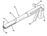
- the surgical training instrument is a linear surgical stapler 10 apparatus comprises a handle 12 at a proximal end and two elongated jaw-like members, an upper jaw 14 and a lower jaw 16 , at a distal end.
- An elongate shaft 18 extends between the handle 12 and the jaws 14 , 16 .
- the elongate shaft 18 houses an actuator shaft (not shown) that is mechanically connected to the handle 12 at the proximal end with gears such that movement of the trigger 20 moves the actuator shaft distally and proximally inside the elongate shaft 18 .
- Such forth and back movement of the actuator shaft permits the distal end of the actuator shaft to ramp in and out of a longitudinal slot 22 in one of the jaw-like members 14 , 16 .
- the longitudinal slot 22 is shown in the upper jaw 14 and advancement of the distal end of the actuator shaft into the longitudinal slot 22 articulates the upper jaw 14 into a closed orientation with respect to the lower jaw 16 .
- the longitudinal slot 22 has a T-shape. Release of the trigger 20 retracts the actuator shaft from the longitudinal slot 22 allowing the upper jaw 14 to open.
- the upper jaw 14 is biased with a spring in the open position. The user can close and open the jaws 14 , 16 by pulling and releasing the trigger 20 until the jaws 14 , 16 are closed to capture simulated tissue at a desired location.
- a complementary longitudinal slot 26 is formed in the lower jaw 16 and the distal end of the actuator shaft slides along the length inside the longitudinal slots 22 , 26 of the closed jaws 14 , 16 .
- the longitudinal slot 26 of the lower jaw 16 is T-shaped. With the jaws 14 , 16 closed, the longitudinal slots 22 , 26 form an I-shape which is complementary in shape to an I-beam of the distal end of the actuator shaft. This configuration assists in keeping the jaws 14 , 16 in the closed configuration.
- the distal end of the actuator shaft includes a cutting element or blade (not shown) that cuts simulated tissue along the longitudinal axis of the jaw 14 , 16 as the actuator shaft is moved distally.
- the trigger 20 is configured to move the actuator distally after release of a safety button such that the trigger 20 can be pulled all way proximally without hindrance.
- the lever 24 is also used to retract the blade by pulling the lever 24 proximally.
- FIGS. 3-4 there is shown another variation for incorporating a cutting element or blade in a surgical training instrument 10 such as a simulated linear stapler 10 or simulated energy-based ligation tool.
- the simulated tool or stapler 28 of FIGS. 3-4 includes movable jaws 30 , 32 similar to the jaws 14 , 16 shown in FIGS. 1-2 .
- Each of the opposing jaws 30 , 32 comprise an opposing surface 34 , 36 , respectively.
- These opposing surfaces 34 , 36 are substantially flat and together provide a compressive force onto simulated tissue material captured between the jaws 30 , 32 when the jaws 30 , 32 are in a closed orientation.
- a cutting element 38 is included in one of the jaws 30 , 32 and is shown in FIG.
- the cutting element 38 is a blade or other protrusion that is sufficiently capable of severing simulated tissue material.
- the cutting element 38 protrudes upwardly from the opposing surface 36 and extends longitudinally along the center line of the lower jaw 32 .
- a slot 40 is included in the opposing surface 34 of the upper jaw 30 directly opposite from the cutting element 38 .
- the slot 40 extends longitudinally along the center line of the upper jaw 30 and is sized and configured to receive at least part of the cutting element 38 .
- the configuration of the cutting element 38 and slot 40 is such that material compressed between the jaws 30 , 32 is severed upon compression or closure of the jaws 30 , 32 .
- the proximal end of the tool 28 comprising a handle and actuators, is not shown in FIGS. 3-4 but are described with respect to FIGS. 1-2 .
- Other variations known to a person having ordinary skill in the art to make the jaws open and close, lock and sever simulated tissue material captured within the jaws 14 , 16 are within the scope of the present invention.
- the jaw-like members 14 , 16 , 30 , 32 articulate relative to each other to open and close to capture material such as simulated tissue material between the jaw-like members 14 , 16 , 30 , 32 .
- the user controls the device 10 from the handle 12 to open and close the jaw-like members 14 , 16 , 30 , 32 and, in general, manipulate and control the device 10 .
- one of the jaw members carries a disposable cartridge containing staplers arranged in two or more rows.
- the other one of the jaw-like members comprises an anvil against which the staples are driven to deform the staple legs.
- Staples are driven out of the cartridge by a camming surface or slider that moves longitudinally against a plurality of laterally positioned pushers that push each staple out of the cartridge individually.
- Surgical staplers typically include a blade that follows the camming surface so as to cut the tissue between the two or more rows of delivered staples.
- the surgical training instrument in the form of a linear stapler 10 does not carry any staples as the staple cartridges are expensive.
- the upper jaw 14 includes a planar opposing surface 15 that is made to resemble the anvil surface of a real stapler.
- the anvil surface is configured to properly deform staples that would exit from openings in the staple cartridge.
- the lower jaw 16 includes a planar opposing surface 17 that resembles a staple cartridge and may comprise an actual staple cartridge from a real linear stapler without the staples. All of the surgical training instruments of the present invention comprise a modified or simulated surgical instrument adapted for training purposes.
- the planar surface 17 of the lower jaw 16 is opposite from the opposing surface 15 of the upper jaw 14 .
- the opposing surface 15 of the upper jaw 14 is marked, textured or embossed to replicate the appearance and position of multiple rows of an anvil surface having a plurality of staple pockets.
- Replicated staple pockets comprising concave indentations are provided in the opposing surface 15 .
- the staple pockets comprise convex protrusions configured to lift and/or carry and transfer ink or other marking compound in order to create realistic markings of staple deployment on one or more sides of the simulated tissue structure.
- a real staple cartridge houses a plurality of staples and includes exit openings in the opposing surface of the lower jaw.
- the opposing surface 17 of the lower jaw 16 is marked, textured or embossed to replicate the appearance and position of multiple rows of staple exit openings.
- the opposing surface 17 of the lower jaw 16 may include small openings that replicate openings through which staples are ejected and exit the cartridge.
- the opposing surface 17 includes raised portions in the location of real staple exit opening configured to impart the other side of the simulated tissue structure with markings and, as such, is configured to lift and/or carry and transfer ink or other marking compound.
- one or more rows of replicated exit openings 42 is provided. Three rows of replicated exit openings 42 on either side of the longitudinal slot 26 are shown in FIGS. 2, 5 and 6 .
- the rows of replicated exit openings 42 are staggered with respect to adjacent rows.
- the opposing surface 15 of the upper jaw 14 includes replicated anvil pockets 44 that are visible in FIG. 5 .
- the replicated anvil pockets 44 are arranged in at least one row on either side of the longitudinal slot 22 of the upper jaw 14 .
- the replicated anvil pockets 44 are aligned directly opposite from the replicated exit openings 42 and are formed in rows that are staggered with respect to adjacent rows. Three rows of replicated anvil pockets 44 on either side of the longitudinal slot 22 are shown in FIG. 5 .
- At least one of the opposing surfaces 15 , 17 includes texture or embossments that are formed in plastic, metal, rubber or other material such that the texture or embossments in the location of replicated exit openings 42 and/or replicated anvil pockets 44 are raised or depressed from the planar opposing surfaces 15 , 17 .
- the upper opposing surface 15 and the lower opposing surface 17 are configured to compress material, typically material simulating tissue such as silicone, plastic, thermoplastic elastomer or other material, between the jaws 14 , 16 , 30 , 32 .
- material typically material simulating tissue such as silicone, plastic, thermoplastic elastomer or other material
- the texturing or embossment on one or more of the opposing surfaces 15 , 17 , 34 , 36 leave a three-dimensional imprint in the simulated tissue material that mimics real rows of staples delivered in the simulated tissue material that is visible to the user.
- these raised locations may be provided with ink, dye, or other material or marking compound having transferable color.
- At least one of the opposing surfaces 15 , 17 , 34 , 36 of the upper and lower jaws 14 , 16 , 30 , 32 respectively, may be compressed upon an inking pad prior to contact with simulated tissue material. This action will deposit a marking fluid, ink, dye, paste or powder upon the marking element comprising textured surfaces 15 , 17 , 34 , 36 and when the jaws 14 , 16 , 30 , 32 are closed down upon simulated tissue material, the markings will be transferred to the simulated tissue material leaving behind a realistic staple-like imprint.
- An inking pad may also be integrally formed within at least one of the jaws 14 , 16 , 30 , 32 containing a carrier for dye, ink or other marking compound.
- the carrier may comprise an ink cartridge, sponge or inking pad.
- the marking element is not limited to that shown in FIGS. 5-6 and may comprise any surface integral or not with the surgical training instrument that is configured to carry a marking compound and to release the marking compound in a pattern upon contact with the outer surface of the simulated tissue structure.
- the embossments would leave a three-dimensional imprint upon the simulated tissue material
- the present invention is not so limited and the embossments, in particular, raised portions of either or both opposing surfaces 15 , 17 , 34 , 36 are configured to leave a two-dimensional deposit of ink or other marking compound on the simulated tissue material without physically deforming the simulated tissue material.
- both a three-dimensional marking and color imprint or transfer of dye and the like upon the outer surface of the simulated tissue structure are within the scope of the present invention.
- an inking pad or sponge is provided.
- the inking pad or sponge is contained within a non-permeable container that is sized and configured to receive and direct a surgical training instrument 10 .
- the container comprises a first portion containing an inking element such as a pad or sponge.
- a second portion of the container receives an instrument in a preferred orientation that facilitates presentation of the surfaces to be treated to the inking element.
- a third portion seals the container when not in use.
- the first portion is generally enlarged relative to the second portion and generally flat so that the marking element is held in a planar orientation.
- the second portion is configured so that an instrument inserted into the second portion approaches the inking element with the surfaces to be inked aligned with the flat surfaces of the inking element.
- the second portion is tubular or obliquely tubular so that the inserted surgical training instrument 10 is inserted with marking structures slightly separated for presentation to the inking element.
- a linear stapler is inserted into the second portion with jaws open or slightly open and moved into the first portion where the jaws are subsequently closed upon the inking element transferring ink, dye or other marking compound to the jaws.
- the inking pad or sponge is a double-sided pad or sponge that transfers ink from two oppositely disposed sides of the pad with one side contacting the first jaw and the other side of the pad contacting the second jaw when the jaws are closed down upon the inking pad.
- the third portion comprises any number of closure elements such as a zip lock, hook-and-loop type fastener or other closing device.
- a kit comprises at least one surgical training instrument and ink pad.
- the kit may additionally comprise at least one simulated tissue structure with or without adhesive according to the present invention.
- a first marking element 46 is provided.
- the first marking element 46 is shaped and configured to slide over the lower jaw 16 .
- the first marking element 46 is sleeve-like having an inner lumen 48 that receives the lower jaw 16 .
- the first marking element 46 includes a planar surface 50 that includes a marking compound and is made of a material, such as a sponge-like material, that will exude the marking compound through specific regions that are arranged upon the marking element 46 , the opposing surface 15 of the upper jaw 14 , the opposing surface 17 of the lower jaw 16 , or a planar surface 52 of a second marking element 54 if one is employed.
- a second marking element 54 is configured with a lumen 56 to slide over and fit on the upper jaw 14 in the same sleeve-like fashion as the first marking element 46 as shown in FIG. 5 .
- the second marking element 54 may also be provided with a marking compound and configured to exude the marking compound through specific regions. These specific regions may be arranged upon the marking element 54 , the opposing surface 17 of the lower jaw 16 , the opposing surface 15 of the upper jaw 14 or on the planar surface 50 of a first marking element 46 if a first marking element 46 is employed. It is clear that at least one of the jaws 14 , 16 is provided with a marking element.
- FIG. 6 illustrates two marking elements 46 , 54 placed on the upper and lower jaws 14 , 16 , respectively.
- the marking elements 46 , 54 may be removed and replaced in simulation of the replacement of staple cartridges in an actual surgical linear stapler. As mentioned above, multiple staple cartridges may be necessary to cut across a larger section of tissue and used staple cartridges would have to be removed and replaced with a new staple cartridge for continued firing.
- the present invention advantageously provides at least one marking element that would provide the practitioner using the simulated tissue stapler 10 with the same action as required in using a real linear surgical stapler.
- the first marking element 46 that is placed over the lower jaw 16 that simulates the staple cartridge would be the marking element 46 that would be removed and replaced.
- that marking element 46 would be the ink-bearing element.
- the marking elements 46 , 54 are configured to create a resulting visual impression in the simulated tissue or organ that mimics the end result of real stapling at the surgical site in color, texture and visual impression upon the user including the use of red or other-colored marking compound.
- the present invention additionally comprises simulated tissue material and structures configured to function with the surgical training instrument 10 such as a linear stapler or other simulated tool, ligation, occluding, or cutting instrument 10 .
- Simulated tissue material and structures include any adjacent tissue surfaces, body conduits, arteries, veins and hollow organs made of elastomeric materials such as silicone, vinyl, polyurethane or any other polymer. Some of the adjacent tissue surfaces, body conduits, arteries, veins, hollow organs, or other tissue structure may include other material or are made solely from other material such as nylon, polyester, cotton or the like.
- the inner surfaces or portions of inner surfaces, or portions of other surfaces such as adjacent surfaces of the simulated tissue material or structure such as conduits, arteries, veins, and hollow organs are supplied with or coated with an adhesive such as a pressure-sensitive adhesive or contact adhesive.
- the simulated tissue structure includes an outer surface, a first inner surface and a second inner surface.
- the second inner surface is adjacent to and faces, at least partially, the first inner surface.
- the adhesive is located on at least one of the first inner surface and the second inner surface. The adhesive remains adhered to one of the inner surfaces and not adhered to the other of the inner surface.
- the cavity or gap in the simulated tissue structure between the first inner surface and the second inner surface is not closed until the surgical training instrument is acted upon the region with adhesive.
- the adhesive is activated to adhere the first inner surface to the second inner surface.
- the jaws bring the inner surfaces together, compress them and adheres the first inner surface to the second inner surface in the location of compression and adhesive simulating surgical occlusion.
- the first inner surface and the second inner surface are located on the inner surface of the lumen.
- the simulated tissue will be compressed between the opposing surfaces 15 , 17 .
- adhesive such as pressure-sensitive adhesive, double-sided tape, or contact adhesive, in particular, in at least the location of surgically desired placement of the simulated tool 10 .
- the inner surfaces or portions of inners surfaces of conduits, arteries, veins and hollow organs that face each other are supplied with or coated with an adhesive such as a pressure-sensitive adhesive or a contact adhesive on at least one of the surfaces.
- a hollow simulated tissue structure includes an inner surface opposite from another inner surface. At least one of these surfaces is coated with adhesive for the purpose of attachment to the other of these surfaces.
- the inner surfaces, supplied with adhesive are forced into occlusion and are adhered together as would tissue that was stapled or welded by a linear surgical stapler, energy-based ligation instrument, or other surgical device.
- the simulated tissue structure is provided with attractive elements instead of adhesive-based elements. Examples of attractive elements include hook-and-loop type fasteners such as VELCRO®, magnets, or the like attached to walls of the simulated tissue structure.
- An example of an adhesive for use in the present invention is Styrene Block Copolymer (SBC) containing rosin or other tackifiers that render a sensitive, tacky surface.
- SBC Styrene Block Copolymer
- Other alternate materials are materials that are formulated to stick to themselves under compression.
- silicone rubber may be compounded or formulated to fuse under pressure in certain conditions.
- An additional material choice comprises a naturally sticky material such as KRATON® gel or the like with or without a non-sticky external coating or external surface.
- a simulated tissue structure 60 is shown in the shape of a hollow organ or body conduit.
- the simulated tissue structure 60 is an elastomeric and/or fabric structure having a wall 62 having an inner surface 68 and an outer surface 70 .
- the simulated tissue structure 60 includes a lumen 64 .
- An occlusive adhesive element 66 is provided on at least a portion of the internal surface or first inner surface along at least a portion of the length of the simulated tissue structure 60 .
- the simulated tissue structure 60 is shown in FIG. 8 in a compressed configuration following a simulated surgical stapling or energy-based procedure showing the adherence of opposing adjacent surfaces along line 72 .
- a surgical training instrument 10 such as a simulated energy-based grasper or dissector 10 is shown adjacent to a simulated tissue structure 60 shown in cross-section defining a lumen 64 .
- the simulated tissue structure 60 is tubular in shape having a circular cross-section resembling a body conduit such as an artery or vein or other hollow organ.
- An inner surface 68 of the simulated tissue structure 60 is provided with adhesive 66 on at least a portion of the inner surface 68 .
- FIG. 9 shows the adhesive 66 placed circumferentially around the inner surface.
- the distal end of the simulated surgical instrument 10 is shown in FIGS. 9-11 .
- the surgical training instrument 10 comprises a pair of opposed jaws 14 , 16 , 30 , 32 as described above.
- the jaws 14 , 16 , 30 , 32 are opened and located adjacent to a surgically desirable location at the simulated tissue structure 60 .
- the handle 12 is manipulated to close the jaws 14 , 16 , 30 , 32 on the simulated tissue structure 60 compressing the simulated tissue structure 60 between the jaws 14 , 16 , 30 , 32 as shown in FIG. 10 .
- the simulated surgical instrument 10 includes a cutting element or blade it is activated to sever the simulated tissue structure in the desired location.
- the simulated tissue structure 10 has an adhesive 66 such as a pressure-sensitive contact adhesive within the lumen 64 so that upon compression and severance, the cut ends appear to have been treated with an energy-based, electro-surgical or electrocautery device or the like with markings, ink-based or otherwise, being imparted to the surface of the simulated tissue structure 60 .
- the jaws 14 , 16 , 30 , 32 are then manipulated at the handle 12 into an open orientation and the severed simulated tissue structure 60 is removed from the instrument 10 as shown in FIG. 11 .
- the simulated tissue structure 60 remains compressed because of the adhesive being activated upon compression or juxtaposition of the adjacent opposing surfaces with at least one surface bearing adhesive for creating a closed lumen 64 as would result from a real occlusive surgical instrument.
- a simulated tissue structure 60 in the shape of a tubular body conduit is shown having a lesion 74 that is to be removed.
- the conduit is elongate and defines a lumen 64 wherein at least a portion of the inner surface of the conduit is provided with adhesive as described above.
- the adhesive may be selectively placed, for example, on either side of the tumor 74 or throughout the entirety of the inside of the lumen 64 .
- a surgical training instrument 10 such as a stapler is shown with jaws 14 , 16 closed upon a portion of the conduit 60 .
- the simulated stapler 10 is operated to simulate the firing of staples by pulling the trigger 20 or advancing the lever 24 to advance a blade. If the surgical training instrument 28 of FIGS.
- the jaws 30 , 32 are closed to a first closed position in which the cutting element 38 does not sever the simulated tissue 60 and then repositioned as needed to the desired location.
- the jaws 30 , 32 are moved to a second closed position in which the simulated tissue is severed by the jaws 30 , 32 coming sufficiently close together to engage the cutting element 38 in one jaw against the opposite jaw.
- the jaws are opened and the simulated instrument 10 is removed leaving behind a plurality of simulated rows 76 , 78 of staples which comprise a pattern of markings that resemble real staples.
- the pattern of markings are from ink, dye or other marking compound carried by the surgical training instrument 10 and imprinted upon or transferred to the outer surface of the simulated tissue structure 60 as shown in FIG. 13 .
- Both sides of the simulated tissue structure 60 is marked in which both jaws are inked. Alternatively, only the side facing the user is marked in which only one of the jaws such as the top jaw is inked.
- Six rows of markings comprising three rows 76 on one side and three rows 78 on another side of the cut 80 are shown in FIG. 13 .
- the markings may include not only color ink placement on the simulated tissue structure 60 , but also, a three dimensional impression or embossment upon the simulated tissue structure 60 that mimic the raised and depressed areas of a real deployment of surgical staples.
- the simulated stapler 10 When the simulated stapler 10 is removed from the conduit 60 , at least one of the marking elements 46 , 54 is removed and replaced to simulate the reloading of a new and real staple cartridge.
- the jaws are inked by compressing the jaws 14 , 16 onto an ink pad to transfer ink to at least one of the jaws.
- the simulated stapler 10 is then reintroduced into the practice area, such as a laparoscopic trainer, and the jaws 14 , 16 are placed in a second location at the other side of the lesion 74 .
- the jaws 14 , 16 are closed down by pulling of the trigger 20 and the simulated stapler 10 is activated again to mimic the firing of staples resulting in the simulated tissue 60 being cut in a second location leaving the similar pattern of ink markings on the simulated tissue 60 described above.
- the simulated tissue structure 60 that is located between the two cuts and containing the lesion 74 is removed. Compression of the simulated tissue structure 60 between the jaws has sealed the open lumen 64 closed in the location of the markings 76 , 78 on either side of the cut 80 and also at the other side of the lesion 74 in the performance of a second cut.
- the simulated tissue structure 60 may be placed inside a laparoscopic trainer 82 such as the one depicted in FIG. 14 and described in co-pending U.S. patent application Ser. No. 13/248,449 entitled “Portable laparoscopic trainer” and filed on Sep. 29, 2011 by Pravong et al. to Applied Medical Resources Corporation and published as U.S. Patent Application Publication No. 2012/0082970, hereby incorporated by reference in its entirety herein.
- Other simulators and/or trainers may be used with the surgical training instruments and simulated tissue structures of the present invention.
- a laparoscopic trainer 82 includes a top cover 84 connected to a base 86 by a plurality of legs 88 spacing the top cover 84 from the base 86 .
- the laparoscopic trainer 82 is configured to mimic the torso of a patient such as the abdominal region.
- the top cover 84 is representative of the anterior surface of the patient and a space defined between the top cover 82 and the base 86 is representative of an interior of the patient or body cavity where organs reside.
- the laparoscopic trainer 82 is a useful tool for teaching, practicing and demonstrating various surgical procedures and their related instruments in simulation of a patient.
- surgical instruments such as the surgical training instrument 10 of the present invention are inserted into the cavity 90 of the laparoscopic trainer 20 through pre-established apertures 92 in the top cover 84 .
- These pre-established apertures 92 may include seals that simulate trocars or may include simulated tissue that simulates the patient's skin and abdominal wall portions.
- Various tools and techniques may be used to penetrate the top cover 84 to perform mock procedures on simulated tissue structures 60 of the present invention placed between the top cover 84 and the base 86 .
- An elongated conduit 60 is shown placed in the cavity 90 of the trainer 82 shown in FIG. 14 .
- the simulated tissue structure 60 When placed inside the cavity 90 of the trainer 82 , the simulated tissue structure 60 is generally obscured from the perspective of the user who can then practice performing surgical techniques laparoscopically by viewing the surgical site indirectly via a video feed displayed on a video monitor 94 .
- the video display monitor 94 is hinged to the top cover 84 and is shown in a closed orientation in FIG. 14 .
- the video monitor 94 is connectable to a variety of visual systems for delivering an image to the monitor 94 .
- a laparoscope inserted through one of the pre-established apertures 92 or a webcam located in the cavity 90 and used to observe the simulated procedure can be connected to the video monitor 94 and/or a mobile computing device to provide an image to the user.
- FIG. 15 illustrates the final end condition of a portion of simulated tissue structure 60 that has the effects of being stapled by a linear stapler 10 described in a procedure shown in FIGS. 12-13 carried out in a laparoscopic trainer 82 or other simulator, for example, and removed.
- the surgical effects include a compressed lumen 64 in the location where two adjacent wall portions 62 A, 62 B are tightly approximated by the closure of jaws.
- the lumen 64 of the conduit 60 is closed and sealed by the adhesive.
- the end condition is replicated or simulated without the use of real staples and what remains are markings from ink or dye and/or embossments or texturing from being compressed by the jaws 14 , 16 .
- three rows of markings 78 resembling three rows of staples are adjacent to the cut line.
- the simulated tissue 60 of the present invention advantageously permits the practice of a leak test to ensure that proper stapling or ligation has taken place.
- a leak test is performed by pumping air or other gas into the conduit 60 at one end and then applying water or other liquid such as saline solution at the cut line and along the markings 78 and observing to see if any bubbles are formed. If bubbles appear, then the practitioner knows that the conduit was not properly sealed by the ligation or stapling instrument.
- adhesive applied to the inside of the lumen may be selectively applied to create the desired passing or failing of the leak test.
- An elastomeric material is used as a body conduit or simulated tissue structure 60 having a lumen 64 .
- an adhesive such as a pressure-sensitive, contact adhesive is deposited or supplied so that upon compression of the elastomeric lumen 64 , the adhesive is fully occluded and activated.
- the opposing walls 62 A, 62 B of the simulated conduit 60 are compressed, sealed and marked in a manner that appears to have been accomplished with a linear stapler employing real surgical staples or other occlusion device, thereby, simulating a surgical procedure.
- a pressure-sensitive contact adhesive or element is placed within the lumen 64 of a simulated body conduit 10 . It may be deposited upon the inner surface 68 using an adhesive applicator 96 or may comprise a wafer or strip layer 98 of double-sided material placed within the lumen 64 .
- a simulated organ 100 resembling a stomach configured for the purposes of practicing a surgical procedure such as a simulated gastric bypass procedure.
- a portion 102 of a stomach 100 is amputated and removed from a body cavity.
- the stomach 100 is representative of other organs upon which similar resection procedures can be practiced.
- the procedure is commonly accomplished using a linear stapler along a prescribed path 104 .
- a synthetic, elastomeric and/or fabric stomach 100 is provided with an occlusive and adherent internal surface such as from an adhesive or adhesive-like material.
- a simulated linear stapler 10 is configured to compress and cut the synthetic stomach 100 along a predetermined path 104 .
- FIG. 19 shows a simulated linear stapler positioned on a simulated stomach 100 .
- the upper and lower jaws 14 , 16 of the simulated stapler 10 close to compress the material and a centrally moving blade cuts through the compressed material leaving a cut 106 and rows of markings 76 , 78 of simulated staple deployment as shown in FIG. 20 .
- the stapler 10 is then removed from the site and a simulated reloading of staple cartridges is performed, for example, by the removal and replacement of one or more marking elements 46 , 54 if they are employed or by re-inking of ink-absorbent material on at least one of the opposing surfaces 15 , 17 , 34 , 36 , 50 , 52 by compressing the jaws onto an ink pad.
- the stapler 10 is again introduced into the simulator or laparoscopic trainer 82 and placed upon the stomach 100 ahead of the cut 106 previously made as shown in FIG. 21 . The action is repeated as shown in FIGS. 22-23 until the stomach 100 is completely divided as shown in FIG.
- the hollow organ 100 comprises two adjacent simulated tissue surfaces that are brought into contact with each other and in the presence of adhesive or adherent material, the adjacent surfaces are glued. If no adhesive or adherent material is present, the compressed adjacent surfaces will not be adhered. Adhesive is selectively placed along one or more of the two adjacent surfaces, placing adhesive where the cut is desired and not locating adhesive where the surgical pathway is unwanted or surgically incorrect.
- the two adjacent surfaces may be pre-adhered along a predetermined pathway that is most desirable for a practitioner to follow in order to achieve a desired clinical outcome in the simulation.
- Adjacent inner surfaces that are pre-adhered are glued together prior to application of the simulated training instrument 10 , in particular, prior to the closure of the jaws onto the simulated tissue structure in the location of the two adjacent surfaces. In such a case, the surgical training instrument 10 does not play a role in adhering the two adjacent surfaces and only severs the pre-adhered surfaces.
- the simulated tissue structure is formed with a pre-adhered portion located along a predetermined pathway.
- the pre-adhered adjacent surfaces define the predetermined pathway 104 for the practitioner to follow.
- the presence of a predetermined pathway that is surgically or clinically significant for the particular procedure is a useful training tool indicating to the practitioner where the cut or stapling should be performed.
- the predetermined pathway of pre-adhered adjacent surfaces is visible to the user.
- the predetermined pathway of pre-adhered adjacent surfaces is only slightly visible to the user.
- the predetermined pathway of pre-adhered adjacent surfaces is not visible to the user.
- the predetermined pathway is molded directly into the simulated tissue structure.
- the predetermined pathway includes a length and a width. The width of the predetermined pathway is at least as wide as the jaws such that when the jaws are placed on the predetermined pathway and the jaws are closed and the blade is activated to create a cut line in the simulated tissue, the simulated tissue structure advantageously remains sealed on either side of the cut line.
- the simulated tissue structure comprises a lumen
- the predetermined pathway divides the lumen into two sections. If the simulated tissue structure is configured as a stomach, the predetermined pathway divides the stomach into two cavities.
Landscapes
- Engineering & Computer Science (AREA)
- Physics & Mathematics (AREA)
- General Physics & Mathematics (AREA)
- Health & Medical Sciences (AREA)
- Mathematical Analysis (AREA)
- Pure & Applied Mathematics (AREA)
- Medical Informatics (AREA)
- Algebra (AREA)
- Computational Mathematics (AREA)
- General Health & Medical Sciences (AREA)
- Chemical & Material Sciences (AREA)
- Mathematical Optimization (AREA)
- Mathematical Physics (AREA)
- Medicinal Chemistry (AREA)
- Business, Economics & Management (AREA)
- Educational Administration (AREA)
- Educational Technology (AREA)
- Theoretical Computer Science (AREA)
- Pulmonology (AREA)
- Radiology & Medical Imaging (AREA)
- Instructional Devices (AREA)
- Surgical Instruments (AREA)
Abstract
Description
Claims (9)
Priority Applications (1)
| Application Number | Priority Date | Filing Date | Title |
|---|---|---|---|
| US13/957,973 US10198965B2 (en) | 2012-08-03 | 2013-08-02 | Simulated stapling and energy based ligation for surgical training |
Applications Claiming Priority (2)
| Application Number | Priority Date | Filing Date | Title |
|---|---|---|---|
| US201261679494P | 2012-08-03 | 2012-08-03 | |
| US13/957,973 US10198965B2 (en) | 2012-08-03 | 2013-08-02 | Simulated stapling and energy based ligation for surgical training |
Publications (2)
| Publication Number | Publication Date |
|---|---|
| US20140038151A1 US20140038151A1 (en) | 2014-02-06 |
| US10198965B2 true US10198965B2 (en) | 2019-02-05 |
Family
ID=49000616
Family Applications (1)
| Application Number | Title | Priority Date | Filing Date |
|---|---|---|---|
| US13/957,973 Expired - Fee Related US10198965B2 (en) | 2012-08-03 | 2013-08-02 | Simulated stapling and energy based ligation for surgical training |
Country Status (7)
| Country | Link |
|---|---|
| US (1) | US10198965B2 (en) |
| EP (1) | EP2880647A1 (en) |
| JP (1) | JP2015525904A (en) |
| KR (1) | KR20150037987A (en) |
| AU (1) | AU2013296222B2 (en) |
| CA (1) | CA2880277A1 (en) |
| WO (1) | WO2014022815A1 (en) |
Cited By (107)
| Publication number | Priority date | Publication date | Assignee | Title |
|---|---|---|---|---|
| US11026713B2 (en) | 2017-10-30 | 2021-06-08 | Cilag Gmbh International | Surgical clip applier configured to store clips in a stored state |
| US11026751B2 (en) | 2017-12-28 | 2021-06-08 | Cilag Gmbh International | Display of alignment of staple cartridge to prior linear staple line |
| US11045591B2 (en) | 2017-12-28 | 2021-06-29 | Cilag Gmbh International | Dual in-series large and small droplet filters |
| US11045197B2 (en) | 2017-10-30 | 2021-06-29 | Cilag Gmbh International | Clip applier comprising a movable clip magazine |
| US11051876B2 (en) | 2017-12-28 | 2021-07-06 | Cilag Gmbh International | Surgical evacuation flow paths |
| US11056244B2 (en) | 2017-12-28 | 2021-07-06 | Cilag Gmbh International | Automated data scaling, alignment, and organizing based on predefined parameters within surgical networks |
| US11058498B2 (en) | 2017-12-28 | 2021-07-13 | Cilag Gmbh International | Cooperative surgical actions for robot-assisted surgical platforms |
| US11076921B2 (en) | 2017-12-28 | 2021-08-03 | Cilag Gmbh International | Adaptive control program updates for surgical hubs |
| US11090047B2 (en) | 2018-03-28 | 2021-08-17 | Cilag Gmbh International | Surgical instrument comprising an adaptive control system |
| US11100631B2 (en) | 2017-12-28 | 2021-08-24 | Cilag Gmbh International | Use of laser light and red-green-blue coloration to determine properties of back scattered light |
| US11096688B2 (en) | 2018-03-28 | 2021-08-24 | Cilag Gmbh International | Rotary driven firing members with different anvil and channel engagement features |
| US11096693B2 (en) | 2017-12-28 | 2021-08-24 | Cilag Gmbh International | Adjustment of staple height of at least one row of staples based on the sensed tissue thickness or force in closing |
| US11114195B2 (en) | 2017-12-28 | 2021-09-07 | Cilag Gmbh International | Surgical instrument with a tissue marking assembly |
| US11129611B2 (en) | 2018-03-28 | 2021-09-28 | Cilag Gmbh International | Surgical staplers with arrangements for maintaining a firing member thereof in a locked configuration unless a compatible cartridge has been installed therein |
| US11132462B2 (en) | 2017-12-28 | 2021-09-28 | Cilag Gmbh International | Data stripping method to interrogate patient records and create anonymized record |
| US11147607B2 (en) | 2017-12-28 | 2021-10-19 | Cilag Gmbh International | Bipolar combination device that automatically adjusts pressure based on energy modality |
| US11160605B2 (en) | 2017-12-28 | 2021-11-02 | Cilag Gmbh International | Surgical evacuation sensing and motor control |
| US11166772B2 (en) | 2017-12-28 | 2021-11-09 | Cilag Gmbh International | Surgical hub coordination of control and communication of operating room devices |
| US11179175B2 (en) | 2017-12-28 | 2021-11-23 | Cilag Gmbh International | Controlling an ultrasonic surgical instrument according to tissue location |
| US11179204B2 (en) | 2017-12-28 | 2021-11-23 | Cilag Gmbh International | Wireless pairing of a surgical device with another device within a sterile surgical field based on the usage and situational awareness of devices |
| US11179208B2 (en) | 2017-12-28 | 2021-11-23 | Cilag Gmbh International | Cloud-based medical analytics for security and authentication trends and reactive measures |
| US11202570B2 (en) | 2017-12-28 | 2021-12-21 | Cilag Gmbh International | Communication hub and storage device for storing parameters and status of a surgical device to be shared with cloud based analytics systems |
| US11207067B2 (en) | 2018-03-28 | 2021-12-28 | Cilag Gmbh International | Surgical stapling device with separate rotary driven closure and firing systems and firing member that engages both jaws while firing |
| US11219453B2 (en) | 2018-03-28 | 2022-01-11 | Cilag Gmbh International | Surgical stapling devices with cartridge compatible closure and firing lockout arrangements |
| US11229436B2 (en) | 2017-10-30 | 2022-01-25 | Cilag Gmbh International | Surgical system comprising a surgical tool and a surgical hub |
| US11234756B2 (en) | 2017-12-28 | 2022-02-01 | Cilag Gmbh International | Powered surgical tool with predefined adjustable control algorithm for controlling end effector parameter |
| US11257589B2 (en) | 2017-12-28 | 2022-02-22 | Cilag Gmbh International | Real-time analysis of comprehensive cost of all instrumentation used in surgery utilizing data fluidity to track instruments through stocking and in-house processes |
| US11253315B2 (en) | 2017-12-28 | 2022-02-22 | Cilag Gmbh International | Increasing radio frequency to create pad-less monopolar loop |
| US11259830B2 (en) | 2018-03-08 | 2022-03-01 | Cilag Gmbh International | Methods for controlling temperature in ultrasonic device |
| US11259806B2 (en) | 2018-03-28 | 2022-03-01 | Cilag Gmbh International | Surgical stapling devices with features for blocking advancement of a camming assembly of an incompatible cartridge installed therein |
| US11259807B2 (en) | 2019-02-19 | 2022-03-01 | Cilag Gmbh International | Staple cartridges with cam surfaces configured to engage primary and secondary portions of a lockout of a surgical stapling device |
| US11266468B2 (en) | 2017-12-28 | 2022-03-08 | Cilag Gmbh International | Cooperative utilization of data derived from secondary sources by intelligent surgical hubs |
| US11273001B2 (en) | 2017-12-28 | 2022-03-15 | Cilag Gmbh International | Surgical hub and modular device response adjustment based on situational awareness |
| US11278281B2 (en) | 2017-12-28 | 2022-03-22 | Cilag Gmbh International | Interactive surgical system |
| US11278280B2 (en) | 2018-03-28 | 2022-03-22 | Cilag Gmbh International | Surgical instrument comprising a jaw closure lockout |
| US11284936B2 (en) | 2017-12-28 | 2022-03-29 | Cilag Gmbh International | Surgical instrument having a flexible electrode |
| US11291510B2 (en) | 2017-10-30 | 2022-04-05 | Cilag Gmbh International | Method of hub communication with surgical instrument systems |
| US11291495B2 (en) | 2017-12-28 | 2022-04-05 | Cilag Gmbh International | Interruption of energy due to inadvertent capacitive coupling |
| US11298148B2 (en) | 2018-03-08 | 2022-04-12 | Cilag Gmbh International | Live time tissue classification using electrical parameters |
| US11308075B2 (en) | 2017-12-28 | 2022-04-19 | Cilag Gmbh International | Surgical network, instrument, and cloud responses based on validation of received dataset and authentication of its source and integrity |
| US11304745B2 (en) | 2017-12-28 | 2022-04-19 | Cilag Gmbh International | Surgical evacuation sensing and display |
| US11304720B2 (en) | 2017-12-28 | 2022-04-19 | Cilag Gmbh International | Activation of energy devices |
| US11304699B2 (en) | 2017-12-28 | 2022-04-19 | Cilag Gmbh International | Method for adaptive control schemes for surgical network control and interaction |
| US11304763B2 (en) | 2017-12-28 | 2022-04-19 | Cilag Gmbh International | Image capturing of the areas outside the abdomen to improve placement and control of a surgical device in use |
| US11311342B2 (en) | 2017-10-30 | 2022-04-26 | Cilag Gmbh International | Method for communicating with surgical instrument systems |
| US11311306B2 (en) | 2017-12-28 | 2022-04-26 | Cilag Gmbh International | Surgical systems for detecting end effector tissue distribution irregularities |
| US11317937B2 (en) | 2018-03-08 | 2022-05-03 | Cilag Gmbh International | Determining the state of an ultrasonic end effector |
| USD950728S1 (en) | 2019-06-25 | 2022-05-03 | Cilag Gmbh International | Surgical staple cartridge |
| US11317919B2 (en) | 2017-10-30 | 2022-05-03 | Cilag Gmbh International | Clip applier comprising a clip crimping system |
| US11317915B2 (en) | 2019-02-19 | 2022-05-03 | Cilag Gmbh International | Universal cartridge based key feature that unlocks multiple lockout arrangements in different surgical staplers |
| US11324557B2 (en) | 2017-12-28 | 2022-05-10 | Cilag Gmbh International | Surgical instrument with a sensing array |
| USD952144S1 (en) | 2019-06-25 | 2022-05-17 | Cilag Gmbh International | Surgical staple cartridge retainer with firing system authentication key |
| US11337746B2 (en) | 2018-03-08 | 2022-05-24 | Cilag Gmbh International | Smart blade and power pulsing |
| US11357503B2 (en) | 2019-02-19 | 2022-06-14 | Cilag Gmbh International | Staple cartridge retainers with frangible retention features and methods of using same |
| US11364075B2 (en) | 2017-12-28 | 2022-06-21 | Cilag Gmbh International | Radio frequency energy device for delivering combined electrical signals |
| US11369377B2 (en) | 2019-02-19 | 2022-06-28 | Cilag Gmbh International | Surgical stapling assembly with cartridge based retainer configured to unlock a firing lockout |
| US11376002B2 (en) | 2017-12-28 | 2022-07-05 | Cilag Gmbh International | Surgical instrument cartridge sensor assemblies |
| US11389164B2 (en) | 2017-12-28 | 2022-07-19 | Cilag Gmbh International | Method of using reinforced flexible circuits with multiple sensors to optimize performance of radio frequency devices |
| US11410259B2 (en) | 2017-12-28 | 2022-08-09 | Cilag Gmbh International | Adaptive control program updates for surgical devices |
| US11419630B2 (en) | 2017-12-28 | 2022-08-23 | Cilag Gmbh International | Surgical system distributed processing |
| US11424027B2 (en) | 2017-12-28 | 2022-08-23 | Cilag Gmbh International | Method for operating surgical instrument systems |
| US11423007B2 (en) | 2017-12-28 | 2022-08-23 | Cilag Gmbh International | Adjustment of device control programs based on stratified contextual data in addition to the data |
| US11419667B2 (en) | 2017-12-28 | 2022-08-23 | Cilag Gmbh International | Ultrasonic energy device which varies pressure applied by clamp arm to provide threshold control pressure at a cut progression location |
| US11432885B2 (en) | 2017-12-28 | 2022-09-06 | Cilag Gmbh International | Sensing arrangements for robot-assisted surgical platforms |
| USD964564S1 (en) | 2019-06-25 | 2022-09-20 | Cilag Gmbh International | Surgical staple cartridge retainer with a closure system authentication key |
| US11446052B2 (en) | 2017-12-28 | 2022-09-20 | Cilag Gmbh International | Variation of radio frequency and ultrasonic power level in cooperation with varying clamp arm pressure to achieve predefined heat flux or power applied to tissue |
| US11464535B2 (en) | 2017-12-28 | 2022-10-11 | Cilag Gmbh International | Detection of end effector emersion in liquid |
| US11464559B2 (en) | 2017-12-28 | 2022-10-11 | Cilag Gmbh International | Estimating state of ultrasonic end effector and control system therefor |
| US11464511B2 (en) | 2019-02-19 | 2022-10-11 | Cilag Gmbh International | Surgical staple cartridges with movable authentication key arrangements |
| US11471156B2 (en) | 2018-03-28 | 2022-10-18 | Cilag Gmbh International | Surgical stapling devices with improved rotary driven closure systems |
| US11504192B2 (en) | 2014-10-30 | 2022-11-22 | Cilag Gmbh International | Method of hub communication with surgical instrument systems |
| US11510741B2 (en) | 2017-10-30 | 2022-11-29 | Cilag Gmbh International | Method for producing a surgical instrument comprising a smart electrical system |
| US11529187B2 (en) | 2017-12-28 | 2022-12-20 | Cilag Gmbh International | Surgical evacuation sensor arrangements |
| US11540855B2 (en) | 2017-12-28 | 2023-01-03 | Cilag Gmbh International | Controlling activation of an ultrasonic surgical instrument according to the presence of tissue |
| US11559307B2 (en) | 2017-12-28 | 2023-01-24 | Cilag Gmbh International | Method of robotic hub communication, detection, and control |
| US11559308B2 (en) | 2017-12-28 | 2023-01-24 | Cilag Gmbh International | Method for smart energy device infrastructure |
| US11564756B2 (en) | 2017-10-30 | 2023-01-31 | Cilag Gmbh International | Method of hub communication with surgical instrument systems |
| US11571234B2 (en) | 2017-12-28 | 2023-02-07 | Cilag Gmbh International | Temperature control of ultrasonic end effector and control system therefor |
| US11576677B2 (en) | 2017-12-28 | 2023-02-14 | Cilag Gmbh International | Method of hub communication, processing, display, and cloud analytics |
| US11589932B2 (en) | 2017-12-28 | 2023-02-28 | Cilag Gmbh International | Usage and technique analysis of surgeon / staff performance against a baseline to optimize device utilization and performance for both current and future procedures |
| US11589888B2 (en) | 2017-12-28 | 2023-02-28 | Cilag Gmbh International | Method for controlling smart energy devices |
| US11601371B2 (en) | 2017-12-28 | 2023-03-07 | Cilag Gmbh International | Surgical network determination of prioritization of communication, interaction, or processing based on system or device needs |
| US11596291B2 (en) | 2017-12-28 | 2023-03-07 | Cilag Gmbh International | Method of compressing tissue within a stapling device and simultaneously displaying of the location of the tissue within the jaws |
| US11602393B2 (en) | 2017-12-28 | 2023-03-14 | Cilag Gmbh International | Surgical evacuation sensing and generator control |
| US11612444B2 (en) | 2017-12-28 | 2023-03-28 | Cilag Gmbh International | Adjustment of a surgical device function based on situational awareness |
| US11659023B2 (en) | 2017-12-28 | 2023-05-23 | Cilag Gmbh International | Method of hub communication |
| US11666331B2 (en) | 2017-12-28 | 2023-06-06 | Cilag Gmbh International | Systems for detecting proximity of surgical end effector to cancerous tissue |
| US11696760B2 (en) | 2017-12-28 | 2023-07-11 | Cilag Gmbh International | Safety systems for smart powered surgical stapling |
| US11744604B2 (en) | 2017-12-28 | 2023-09-05 | Cilag Gmbh International | Surgical instrument with a hardware-only control circuit |
| US11771487B2 (en) | 2017-12-28 | 2023-10-03 | Cilag Gmbh International | Mechanisms for controlling different electromechanical systems of an electrosurgical instrument |
| US11786245B2 (en) | 2017-12-28 | 2023-10-17 | Cilag Gmbh International | Surgical systems with prioritized data transmission capabilities |
| US11786251B2 (en) | 2017-12-28 | 2023-10-17 | Cilag Gmbh International | Method for adaptive control schemes for surgical network control and interaction |
| US11801098B2 (en) | 2017-10-30 | 2023-10-31 | Cilag Gmbh International | Method of hub communication with surgical instrument systems |
| US11818052B2 (en) | 2017-12-28 | 2023-11-14 | Cilag Gmbh International | Surgical network determination of prioritization of communication, interaction, or processing based on system or device needs |
| US11832899B2 (en) | 2017-12-28 | 2023-12-05 | Cilag Gmbh International | Surgical systems with autonomously adjustable control programs |
| US11832840B2 (en) | 2017-12-28 | 2023-12-05 | Cilag Gmbh International | Surgical instrument having a flexible circuit |
| US11857152B2 (en) | 2017-12-28 | 2024-01-02 | Cilag Gmbh International | Surgical hub spatial awareness to determine devices in operating theater |
| US11864728B2 (en) | 2017-12-28 | 2024-01-09 | Cilag Gmbh International | Characterization of tissue irregularities through the use of mono-chromatic light refractivity |
| US11871901B2 (en) | 2012-05-20 | 2024-01-16 | Cilag Gmbh International | Method for situational awareness for surgical network or surgical network connected device capable of adjusting function based on a sensed situation or usage |
| US11890065B2 (en) | 2017-12-28 | 2024-02-06 | Cilag Gmbh International | Surgical system to limit displacement |
| US11896443B2 (en) | 2017-12-28 | 2024-02-13 | Cilag Gmbh International | Control of a surgical system through a surgical barrier |
| US11896322B2 (en) | 2017-12-28 | 2024-02-13 | Cilag Gmbh International | Sensing the patient position and contact utilizing the mono-polar return pad electrode to provide situational awareness to the hub |
| US11903601B2 (en) | 2017-12-28 | 2024-02-20 | Cilag Gmbh International | Surgical instrument comprising a plurality of drive systems |
| US11903587B2 (en) | 2017-12-28 | 2024-02-20 | Cilag Gmbh International | Adjustment to the surgical stapling control based on situational awareness |
| US11911045B2 (en) | 2017-10-30 | 2024-02-27 | Cllag GmbH International | Method for operating a powered articulating multi-clip applier |
| US11937769B2 (en) | 2017-12-28 | 2024-03-26 | Cilag Gmbh International | Method of hub communication, processing, storage and display |
| US11969216B2 (en) | 2017-12-28 | 2024-04-30 | Cilag Gmbh International | Surgical network recommendations from real time analysis of procedure variables against a baseline highlighting differences from the optimal solution |
Families Citing this family (34)
| Publication number | Priority date | Publication date | Assignee | Title |
|---|---|---|---|---|
| WO2012044753A2 (en) | 2010-10-01 | 2012-04-05 | Applied Medical Resources Corporation | Portable laparoscopic trainer |
| KR101963610B1 (en) | 2011-10-21 | 2019-03-29 | 어플라이드 메디컬 리소시스 코포레이션 | Simulated tissue structure for surgical training |
| US8961190B2 (en) | 2011-12-20 | 2015-02-24 | Applied Medical Resources Corporation | Advanced surgical simulation |
| JP2015525904A (en) | 2012-08-03 | 2015-09-07 | アプライド メディカル リソーシーズ コーポレイション | Simulated stapling and energy-based ligation for surgical training |
| KR102105979B1 (en) | 2012-09-26 | 2020-05-04 | 어플라이드 메디컬 리소시스 코포레이션 | Surgical training model for laparoscopic procedures |
| CA3159450A1 (en) | 2012-09-27 | 2014-04-03 | Applied Medical Resources Corporation | Surgical training model for laparoscopic procedures |
| US10679520B2 (en) | 2012-09-27 | 2020-06-09 | Applied Medical Resources Corporation | Surgical training model for laparoscopic procedures |
| EP3846151B1 (en) | 2012-09-27 | 2023-11-29 | Applied Medical Resources Corporation | Surgical training model for laparoscopic procedures |
| KR20150063143A (en) | 2012-09-28 | 2015-06-08 | 어플라이드 메디컬 리소시스 코포레이션 | Surgical training model for laparoscopic procedures |
| EP2901438B1 (en) | 2012-09-28 | 2019-02-06 | Applied Medical Resources Corporation | Surgical training model for transluminal laparoscopic procedures |
| JP6482478B2 (en) | 2013-03-01 | 2019-03-13 | アプライド メディカル リソーシーズ コーポレイション | Surgical simulation system and method |
| CA2912069C (en) | 2013-05-15 | 2022-01-11 | Applied Medical Resources Corporation | Hernia model |
| EP3301662B1 (en) | 2013-06-18 | 2023-06-07 | Applied Medical Resources Corporation | Gallbladder model |
| KR102239454B1 (en) | 2013-07-24 | 2021-04-13 | 어플라이드 메디컬 리소시스 코포레이션 | First entry model |
| US10198966B2 (en) | 2013-07-24 | 2019-02-05 | Applied Medical Resources Corporation | Advanced first entry model for surgical simulation |
| AU2015235994B2 (en) * | 2014-03-26 | 2019-11-21 | Applied Medical Resources Corporation | Simulated dissectible tissue |
| EP3218892B1 (en) | 2014-11-13 | 2019-10-23 | Applied Medical Resources Corporation | Simulated tissue models and methods |
| KR20230143198A (en) | 2015-02-19 | 2023-10-11 | 어플라이드 메디컬 리소시스 코포레이션 | Simulated tissue structures and methods |
| US10172670B2 (en) * | 2015-03-16 | 2019-01-08 | Ethicon Llc | Flexible neck for surgical instruments |
| KR20160131694A (en) | 2015-05-08 | 2016-11-16 | 주식회사 엔도비전 | Ligation device |
| KR20180008417A (en) | 2015-05-14 | 2018-01-24 | 어플라이드 메디컬 리소시스 코포레이션 | Synthetic tissue structure for electrosurgical training and simulation |
| WO2016201085A1 (en) | 2015-06-09 | 2016-12-15 | Applied Medical Resources Corporation | Hysterectomy model |
| AU2016291726B2 (en) | 2015-07-16 | 2022-02-03 | Applied Medical Resources Corporation | Simulated dissectable tissue |
| US10490105B2 (en) | 2015-07-22 | 2019-11-26 | Applied Medical Resources Corporation | Appendectomy model |
| EP4300467A3 (en) | 2015-10-02 | 2024-04-03 | Applied Medical Resources Corporation | Hysterectomy model |
| US10706743B2 (en) | 2015-11-20 | 2020-07-07 | Applied Medical Resources Corporation | Simulated dissectible tissue |
| WO2018005301A1 (en) | 2016-06-27 | 2018-01-04 | Applied Medical Resources Corporation | Simulated abdominal wall |
| EP3583589A1 (en) | 2017-02-14 | 2019-12-25 | Applied Medical Resources Corporation | Laparoscopic training system |
| US10847057B2 (en) | 2017-02-23 | 2020-11-24 | Applied Medical Resources Corporation | Synthetic tissue structures for electrosurgical training and simulation |
| BR112020011783A2 (en) * | 2017-12-28 | 2021-02-02 | Ethicon Llc | surgical instrument with tissue marking set |
| US20220000573A1 (en) * | 2018-11-27 | 2022-01-06 | Covidien Lp | Haptic feedback device for surgical instruments and robotic surgical systems |
| US11547411B2 (en) * | 2019-02-22 | 2023-01-10 | Covidien Lp | Anastomosis wound protector |
| US20220015856A1 (en) * | 2020-07-14 | 2022-01-20 | Covidien Lp | Surgical stapler with illumination |
| CN113160676B (en) * | 2021-01-06 | 2022-11-11 | 浙江省人民医院 | Operation training model, weight-reducing metabolism operation training model and training method |
Citations (413)
| Publication number | Priority date | Publication date | Assignee | Title |
|---|---|---|---|---|
| US184573A (en) | 1876-11-21 | Improvement in gas-cocks | ||
| US2127774A (en) | 1936-04-27 | 1938-08-23 | Jacobs Julian Bay | Apparatus for teaching obstetrics |
| US2284888A (en) | 1941-04-14 | 1942-06-02 | Arc Diaphragm & Drug Co | Demonstrating device for vaginal diaphragms |
| US2324702A (en) | 1938-11-30 | 1943-07-20 | Karl F Hoffmann | Surgical simulacra and process of preparing same |
| US2345489A (en) | 1943-04-10 | 1944-03-28 | Frederic P Lord | Anatomical model |
| US2495568A (en) | 1948-12-30 | 1950-01-24 | Holland Rantos Company Inc | Clinical model |
| US3766666A (en) | 1971-10-13 | 1973-10-23 | Robins Co Inc A H | Uterine simulator trainer |
| US3775865A (en) | 1972-07-24 | 1973-12-04 | R Rowan | Simulator for teaching suturing techniques |
| US3789518A (en) | 1972-04-12 | 1974-02-05 | Weatherby Nasco Inc | Simulated human limb |
| US3921311A (en) | 1975-01-06 | 1975-11-25 | Pathfinder Fund | Clinical demonstration model |
| US3991490A (en) | 1973-04-09 | 1976-11-16 | Markman H David | Teaching aid for sigmoidoscope and the like |
| US4001951A (en) | 1975-03-25 | 1977-01-11 | Fasse Wolfgang G | Breast cancer detection training device |
| US4001952A (en) | 1975-10-24 | 1977-01-11 | Kleppinger Trygve M | Hysteroscopy teaching aid |
| US4321047A (en) | 1980-06-05 | 1982-03-23 | Bradley Landis | Simulator and process for teaching surgical knot tying techniques |
| US4323350A (en) | 1980-09-22 | 1982-04-06 | Bowden Jr Robert L | Anatomical model |
| US4332569A (en) | 1981-03-16 | 1982-06-01 | University Of Kentucky Research Foundation | Instructional device for use of a bronchoscope |
| US4371345A (en) | 1980-10-17 | 1983-02-01 | National Research Development Corporation | Multi-dimensional display equipment |
| US4386917A (en) | 1981-09-16 | 1983-06-07 | Forrest Leonard E | Suturing training device and method |
| US4459113A (en) | 1981-08-31 | 1984-07-10 | Boscaro Gatti Antonino E | Device for specializing in the use of endoscopes |
| US4481001A (en) * | 1983-05-26 | 1984-11-06 | Collagen Corporation | Human skin model for intradermal injection demonstration or training |
| US4596528A (en) | 1984-07-02 | 1986-06-24 | Lewis Leonard A | Simulated skin and method |
| US4726772A (en) | 1986-12-01 | 1988-02-23 | Kurt Amplatz | Medical simulator |
| US4737109A (en) | 1987-03-03 | 1988-04-12 | Abramson Daniel J | Breast cancer detection training device |
| US4789340A (en) | 1987-08-18 | 1988-12-06 | Zikria Bashir A | Surgical student teaching aid |
| US4832978A (en) | 1987-04-24 | 1989-05-23 | Lesser Jary M | Simulated connective tissue for construction of models and prostheses |
| US4867686A (en) | 1989-02-09 | 1989-09-19 | Goldstein Mark K | Breast cancer detection model and method for using same |
| US4907973A (en) | 1988-11-14 | 1990-03-13 | Hon David C | Expert system simulator for modeling realistic internal environments and performance |
| US4938696A (en) | 1989-07-25 | 1990-07-03 | Foster-Pickard International, Inc. | Model demonstrating human organ systems |
| US4940412A (en) | 1987-12-08 | 1990-07-10 | Elscint Ltd. | Method of manufacturing anatomical models |
| US5061187A (en) | 1990-04-12 | 1991-10-29 | Ravinder Jerath | Ultrasound training apparatus |
| US5083962A (en) | 1987-08-24 | 1992-01-28 | Pracas Victor M | Doll capable of bodily functions |
| US5104328A (en) | 1990-04-18 | 1992-04-14 | Lounsbury Katherine L | Anatomical model |
| DE4105892A1 (en) | 1991-02-14 | 1992-08-27 | Arnold Dipl Ing Dr Med Pier | Simulation training appts. for surgery - comprises sealed housing and cover releasably connected to housing, excess pressure being formable in housing, and having flat insert in cover top side opening |
| US5149270A (en) | 1990-10-29 | 1992-09-22 | Mckeown M J | Apparatus for practicing surgical procedures |
| US5180308A (en) | 1992-01-06 | 1993-01-19 | Garito Jon C | Medical demonstration model |
| US5230630A (en) | 1992-07-20 | 1993-07-27 | Richard Burgett | Suture training device |
| FR2691826A1 (en) | 1992-06-01 | 1993-12-03 | Allal Hossein | Coelio-surgery simulator for teaching and training of surgeons - uses semi-rigid envelope in form of human body, with coeloscope connected to video camera and to video screen |
| US5273435A (en) | 1992-07-16 | 1993-12-28 | The Mcw Research Foundation, Inc. | Tumor localization phantom |
| WO1994006109A1 (en) | 1992-09-07 | 1994-03-17 | Diethelm Wallwiener | Medical training apparatus |
| US5295694A (en) | 1992-10-27 | 1994-03-22 | Levin John M | Laparoscopic surgery simulating game |
| US5310348A (en) | 1992-10-09 | 1994-05-10 | United States Surgical Corporation | Suture demonstration portfolio |
| US5318448A (en) | 1992-01-06 | 1994-06-07 | Garito Jon C | Demonstration model for gynecological procedure |
| US5320537A (en) | 1993-03-16 | 1994-06-14 | Triangle Research And Development Corporation | Microsurgical training apparatus |
| US5358408A (en) | 1993-03-09 | 1994-10-25 | Marelyn Medina | Tissue specimen suspension device |
| US5368487A (en) | 1992-07-31 | 1994-11-29 | Medina; Marelyn | Laparoscopic training device and method of use |
| US5380207A (en) | 1993-12-27 | 1995-01-10 | Siepser; Steven B. | Slip suture practice kit |
| US5403191A (en) | 1991-10-21 | 1995-04-04 | Tuason; Leo B. | Laparoscopic surgery simulator and method of use |
| US5425731A (en) | 1991-04-05 | 1995-06-20 | Metcal, Inc. | Instrument for cutting, coagulating and ablating tissue |
| US5425644A (en) | 1993-05-13 | 1995-06-20 | Gerhard Szinicz | Surgical training apparatus and method |
| DE4414832A1 (en) | 1994-04-28 | 1995-11-02 | Laerdal Asmund S As | Teaching model for practising blood taking or injection of blood vessels |
| US5472345A (en) | 1993-04-14 | 1995-12-05 | Gaumard Scientific Company, Inc. | Gynecological simulator |
| US5518407A (en) | 1993-11-02 | 1996-05-21 | Greenfield; Cathy L. | Anatomically correct artificial organ replicas for use as teaching aids |
| US5518406A (en) | 1993-11-24 | 1996-05-21 | Waters; Tammie C. | Percutaneous endoscopic gastrostomy teaching device |
| US5520633A (en) | 1991-01-03 | 1996-05-28 | Costin; John A. | Computer controlled smart phacoemulsification method and apparatus |
| US5541304A (en) | 1994-05-02 | 1996-07-30 | Hercules Incorporated | Crosslinked hydrogel compositions with improved mechanical performance |
| WO1996042076A1 (en) | 1995-06-09 | 1996-12-27 | Simulab Corporation | Anatomical simulator for videoendoscopic surgical training |
| US5720742A (en) | 1994-10-11 | 1998-02-24 | Zacharias; Jaime | Controller and actuating system for surgical instrument |
| US5722836A (en) | 1996-05-21 | 1998-03-03 | Simulab Corporation | Reflected-image videoendoscopic surgical trainer and method of training |
| US5727948A (en) | 1996-09-05 | 1998-03-17 | Jordan; Lynette S. | Syringe injection practice device |
| US5743730A (en) | 1996-05-07 | 1998-04-28 | Clester; Kenneth E. | Dental porcelain shading guide and method of use therefore |
| US5762458A (en) | 1996-02-20 | 1998-06-09 | Computer Motion, Inc. | Method and apparatus for performing minimally invasive cardiac procedures |
| US5769640A (en) | 1992-12-02 | 1998-06-23 | Cybernet Systems Corporation | Method and system for simulating medical procedures including virtual reality and control method and system for use therein |
| US5775916A (en) | 1992-01-15 | 1998-07-07 | Limbs & Things Limited | Method of making a surgical and/or clinical apparatus |
| US5785531A (en) | 1996-06-06 | 1998-07-28 | Wilson-Cook Medical Incorporated | Cuttable papilla and sphincterotomy training apparatus |
| JPH10211160A (en) | 1997-01-29 | 1998-08-11 | Olympus Optical Co Ltd | Colon endoscope insertion training device |
| US5800178A (en) | 1995-03-29 | 1998-09-01 | Gillio; Robert G. | Virtual surgery input device |
| US5803746A (en) | 1996-01-23 | 1998-09-08 | Medisim Corporation | Body part model and method of making same |
| US5807378A (en) | 1995-06-07 | 1998-09-15 | Sri International | Surgical manipulator for a telerobotic system |
| US5810880A (en) | 1995-06-07 | 1998-09-22 | Sri International | System and method for releasably holding a surgical instrument |
| US5850033A (en) | 1994-09-30 | 1998-12-15 | Mirzeabasov; Timur Akhmedbekovich | Target for simulating biological subjects |
| WO1998058358A1 (en) | 1997-06-19 | 1998-12-23 | Limbs & Things Limited | Clinical and/or surgical training apparatus |
| US5855583A (en) | 1996-02-20 | 1999-01-05 | Computer Motion, Inc. | Method and apparatus for performing minimally invasive cardiac procedures |
| WO1999001074A1 (en) | 1997-07-03 | 1999-01-14 | Neothermia Corporation | Methods and apparatus for therapeutic cauterization of predetermined volumes of biological tissue |
| US5873732A (en) | 1997-09-26 | 1999-02-23 | Hasson; Harrith M. | Apparatus for training for the performance of a medical procedure |
| US5873863A (en) | 1997-08-29 | 1999-02-23 | United States Surgical Corporation | Vascular surgery demonstration/training kit |
| US5908302A (en) | 1998-06-12 | 1999-06-01 | Goldfarb; Michael A. | Inguinal hernia model |
| US5947743A (en) | 1997-09-26 | 1999-09-07 | Hasson; Harrith M. | Apparatus for training for the performance of a medical procedure |
| WO2000036577A1 (en) | 1998-12-14 | 2000-06-22 | Pharmabotics Limited | Simulated body tissue |
| US6083008A (en) | 1997-09-01 | 2000-07-04 | Agency Of Industrial Science & Technology, Ministry Of International Trade & Industry | Optical phantom of living body and method for producing it |
| EP1024173A1 (en) | 1999-01-14 | 2000-08-02 | Dow Corning Corporation | Silicone gel composition and silicone gel produced therefrom |
| US6113395A (en) | 1998-08-18 | 2000-09-05 | Hon; David C. | Selectable instruments with homing devices for haptic virtual reality medical simulation |
| DE19716341C2 (en) | 1997-03-19 | 2000-09-21 | Erbe Elektromedizin | Training model, especially torso |
| JP2001005378A (en) | 1999-06-24 | 2001-01-12 | Yasuhisa Koki:Kk | Simulator for training of operation technique |
| CN2421706Y (en) | 2000-04-26 | 2001-02-28 | 佟玉章 | Multifunctional exerciser for operating tying and suturing |
| US6234804B1 (en) | 1999-03-02 | 2001-05-22 | Peter Yong | Thoracic training model for endoscopic cardiac surgery |
| WO2002038039A2 (en) | 2000-10-23 | 2002-05-16 | Toly Christopher C | Human surgical trainer and methods for training |
| US6398557B1 (en) | 1999-09-17 | 2002-06-04 | The University Of Iowa Research Foundation | Devices, methods and kits for training in surgical techniques |
| US6474993B1 (en) | 1996-12-04 | 2002-11-05 | Erbe Elektromedizin Gmbh | Artificial tissue |
| US20020165541A1 (en) * | 2001-04-20 | 2002-11-07 | Whitman Michael P. | Bipolar or ultrasonic surgical device |
| US20020168619A1 (en) | 1999-12-29 | 2002-11-14 | Provenza Thadeu Rezende | Simulator device for human feminine mammary gland |
| US6485308B1 (en) | 2001-07-09 | 2002-11-26 | Mark K. Goldstein | Training aid for needle biopsy |
| US6488507B1 (en) | 1999-11-29 | 2002-12-03 | Ethicon, Inc. | Portable surgical trainer |
| US6497902B1 (en) | 1999-12-01 | 2002-12-24 | The Regents Of The University Of Michigan | Ionically crosslinked hydrogels with adjustable gelation time |
| US6511325B1 (en) | 1998-05-04 | 2003-01-28 | Advanced Research & Technology Institute | Aortic stent-graft calibration and training model |
| US6517354B1 (en) | 2000-11-17 | 2003-02-11 | David Levy | Medical simulation apparatus and related method |
| US20030031993A1 (en) | 1999-08-30 | 2003-02-13 | Carla Pugh | Medical examination teaching and measurement system |
| US20030091967A1 (en) | 2000-04-12 | 2003-05-15 | Edna Chosack | Endoscopic tutorial system for urology |
| US20030176770A1 (en) | 2000-03-16 | 2003-09-18 | Merril Gregory L. | System and method for controlling force applied to and manipulation of medical instruments |
| MXPA02004422A (en) | 2002-05-03 | 2003-11-07 | Univ Mexico Nacional Autonoma | Training device for surgery of minimal invasion. |
| US6654000B2 (en) | 1994-07-14 | 2003-11-25 | Immersion Corporation | Physically realistic computer simulation of medical procedures |
| US6659776B1 (en) | 2000-12-28 | 2003-12-09 | 3-D Technical Services, Inc. | Portable laparoscopic trainer |
| US20040005423A1 (en) | 2000-05-12 | 2004-01-08 | Dalton Paul D. | Method of producing structures using centrifugal forces |
| WO2004032095A1 (en) | 2002-10-07 | 2004-04-15 | Xitact S.A. | Interactive medical training system and method |
| US6773263B2 (en) | 2001-10-09 | 2004-08-10 | Robert J. Nicholls | Medical simulator |
| WO2004082486A1 (en) | 2003-03-18 | 2004-09-30 | Anke Gasche | Apparatus and method for colonoscopic appendectomy |
| US6820025B2 (en) | 2000-10-30 | 2004-11-16 | The United States Of America As Represented By The Secretary Of The Navy | Method and apparatus for motion tracking of an articulated rigid body |
| US20040248072A1 (en) | 2001-10-02 | 2004-12-09 | Gray Roger Leslie | Method and apparatus for simulation of an endoscopy operation |
| US20050008997A1 (en) | 2003-07-08 | 2005-01-13 | Mayo Foundation For Medical Education And Research | Portable endoscopic training and research device and methods of use |
| US20050026125A1 (en) | 2000-10-23 | 2005-02-03 | Toly Christopher C. | Simulated anatomical structures incorporating an embedded image layer |
| US6854976B1 (en) | 2002-11-02 | 2005-02-15 | John S. Suhr | Breast model teaching aid and method |
| US6857878B1 (en) | 1998-01-26 | 2005-02-22 | Simbionix Ltd. | Endoscopic tutorial system |
| US6866514B2 (en) | 2003-01-31 | 2005-03-15 | Von Enterprises, Inc. | Gel electrophoresis training aid and training kit |
| US20050084833A1 (en) | 2002-05-10 | 2005-04-21 | Gerard Lacey | Surgical training simulator |
| US6887082B2 (en) | 2000-08-23 | 2005-05-03 | The Royal Alexandra Hospital For Children | Laparoscopic trainer |
| US20050131390A1 (en) | 2002-04-25 | 2005-06-16 | Russell Heinrich | Surgical instruments including mems devices |
| US20050142525A1 (en) | 2003-03-10 | 2005-06-30 | Stephane Cotin | Surgical training system for laparoscopic procedures |
| WO2005071639A1 (en) | 2004-01-09 | 2005-08-04 | Board Of Regents, The University Of Texas System | Models imitating internal organs and the real anatomy |
| US6929481B1 (en) | 1996-09-04 | 2005-08-16 | Immersion Medical, Inc. | Interface device and method for interfacing instruments to medical procedure simulation systems |
| US20050196740A1 (en) | 2004-03-08 | 2005-09-08 | Olympus Corporation | Simulator system and training method for endoscopic manipulation using simulator |
| US20050196739A1 (en) | 2004-03-08 | 2005-09-08 | Olympus Corporation | Endoscopic simulator system and training method for endoscopic manipulation using endoscopic simulator |
| US6950025B1 (en) | 2002-05-17 | 2005-09-27 | Li Nguyen | Medical surgery safety device |
| US20050214727A1 (en) | 2004-03-08 | 2005-09-29 | The Johns Hopkins University | Device and method for medical training and evaluation |
| US6960617B2 (en) | 2002-04-22 | 2005-11-01 | Purdue Research Foundation | Hydrogels having enhanced elasticity and mechanical strength properties |
| CN2751372Y (en) | 2004-12-15 | 2006-01-11 | 武彪 | Laparoscope simulated training table |
| US6997719B2 (en) | 2002-06-26 | 2006-02-14 | Ethicon, Inc. | Training model for endoscopic vessel harvesting |
| US7008232B2 (en) | 2001-09-29 | 2006-03-07 | Friedhelm Brassel | Method for producing a modeling system for vessel deformations |
| US7018327B1 (en) | 2002-08-27 | 2006-03-28 | Conti James C | Test apparatus providing pulsatile flow service for test samples |
| US7056123B2 (en) | 2001-07-16 | 2006-06-06 | Immersion Corporation | Interface apparatus with cable-driven force feedback and grounded actuators |
| US7080984B1 (en) | 2002-04-29 | 2006-07-25 | Bonnie Cohen | Simulated disposable foreskin for training surgical procedure of infant circumcision |
| WO2006083963A2 (en) | 2005-02-03 | 2006-08-10 | Christopher Sakezles | Models and methods of using same for testing medical devices |
| US20060252019A1 (en) | 2005-05-06 | 2006-11-09 | David Burkitt | Knot tying training apparatus |
| US20060275741A1 (en) | 2005-06-02 | 2006-12-07 | Depuy Spine, Inc. | Spine simulator system |
| US20070077544A1 (en) | 2005-06-16 | 2007-04-05 | Gottfried Lemperle | Life-like anatomic feature for testing injection of soft tissue fillers |
| US20070078484A1 (en) | 2005-10-03 | 2007-04-05 | Joseph Talarico | Gentle touch surgical instrument and method of using same |
| US20070074584A1 (en) | 2005-10-03 | 2007-04-05 | Joseph Talarico | Gentle touch surgical instrument and method of using same |
| CN2909427Y (en) | 2006-03-21 | 2007-06-06 | 南方医科大学珠江医院 | Surgical basic skill training box |
| WO2007068360A1 (en) | 2005-12-13 | 2007-06-21 | Erbe Elektromedizin Gmbh | Training model for the endoscopic investigation and treatment of hollow organs |
| US20070148626A1 (en) | 2003-10-16 | 2007-06-28 | Seiichi Ikeda | Three-dimensional model |
| US20070166682A1 (en) | 2003-01-22 | 2007-07-19 | Realsim Systems, Llc. | Medical training apparatus |
| US20070179528A1 (en) * | 2006-02-02 | 2007-08-02 | Soltz Michael A | Mechanically tuned buttress material to assist with proper formation of surgical element in diseased tissue |
| US7255565B2 (en) | 2004-03-15 | 2007-08-14 | Brian Keegan | Anthropomorphic phantoms and method |
| US20070197895A1 (en) | 2006-02-17 | 2007-08-23 | Sdgi Holdings, Inc. | Surgical instrument to assess tissue characteristics |
| US7269532B2 (en) | 2002-04-05 | 2007-09-11 | Commissariat A L'energie Atomique | Device and method for measuring orientation of a solid with measurement correction means |
| US7272766B2 (en) | 2005-04-04 | 2007-09-18 | Christopher Sakezles | Method of making tissue simulating analog materials and models made from same |
| US20070225734A1 (en) | 2006-03-22 | 2007-09-27 | Minos Medical | Systems and methods for less invasive resolution of maladies of tissue including the appendix, gall bladder, and hemorrhoids |
| US7300450B2 (en) | 2001-09-03 | 2007-11-27 | Vleugels Holding B.V. | Surgical instrument |
| US20070275359A1 (en) | 2004-06-22 | 2007-11-29 | Rotnes Jan S | Kit, operating element and haptic device for use in surgical simulation systems |
| US20080032273A1 (en) | 2006-06-21 | 2008-02-07 | Boston Scientific Scimed, Inc. | Anatomical model |
| US20080032272A1 (en) | 2006-08-01 | 2008-02-07 | Palakodeti Ratna K | Surgery Practice Kit |
| WO2008021720A2 (en) | 2006-08-14 | 2008-02-21 | Artann Laboratories, Inc. | Human tissue phantoms and methods for manufacturing thereof |
| US20080064017A1 (en) | 2006-08-29 | 2008-03-13 | Grundmeyer Ramond Iii | Suture training device |
| US20080076101A1 (en) | 2006-05-12 | 2008-03-27 | Abbott Laboratories | Forming vascular diseases within anatomical models |
| US20080097501A1 (en) | 2006-06-22 | 2008-04-24 | Tyco Healthcare Group Lp | Ultrasonic probe deflection sensor |
| US7364582B2 (en) | 2003-10-30 | 2008-04-29 | Cambridge Endoscopic Devices, Inc. | Surgical instrument |
| US20080108869A1 (en) | 2006-10-20 | 2008-05-08 | Femsuite Llc | Optical surgical device and methods of use |
| US20080114381A1 (en) * | 2006-11-10 | 2008-05-15 | Voegele James W | Form in place fasteners |
| US20080188948A1 (en) | 2007-02-05 | 2008-08-07 | Flatt Terry J | Liner system and liner for prosthetics and method for using and making |
| US7427199B2 (en) | 2005-02-03 | 2008-09-23 | Christopher Sakezles | Models and methods of using same for testing medical devices |
| US7431189B2 (en) | 2006-08-02 | 2008-10-07 | Ethicon Endo-Surgery, Inc. | Pneumatically powered surgical cutting and fastening instrument with mechanical linkage coupling end effector and trigger motion |
| CN101313842A (en) | 2007-05-29 | 2008-12-03 | 高永东 | Peritoneoscope vermiform appendix ablation lancing retractor |
| US7465168B2 (en) | 2004-09-03 | 2008-12-16 | Birth Injury Prevention, Llc | Birthing simulator |
| US7467075B2 (en) | 2004-12-23 | 2008-12-16 | Covidien Ag | Three-dimensional finite-element code for electrosurgery and thermal ablation simulations |
| FR2917876A1 (en) | 2007-06-22 | 2008-12-26 | Michel Bams | Anatomical pedagogical device for use in e.g. healthcare establishment, has abdominal muscles with strap connecting lower part of sternum to pubis to shorten/extend muscles length for inducing hyperpressure and amplitude limitation |
| WO2009000939A1 (en) | 2007-06-22 | 2008-12-31 | Gmv, S.A. | Laparoscopic surgical simulator |
| US20090068627A1 (en) | 2000-10-23 | 2009-03-12 | Toly Christopher C | Medical physiological simulator including a conductive elastomer layer |
| US20090092651A1 (en) * | 2007-10-05 | 2009-04-09 | Bhavin Shah | Absorbable adhesives and their formulation for use in medical applications |
| US20090142741A1 (en) | 2007-11-29 | 2009-06-04 | Cedars-Sinai Medical Center | Medical training methods and devices |
| US20090143642A1 (en) | 2007-11-29 | 2009-06-04 | Kazuhiko Takahashi | Therapeutic device system and manipulator system |
| US20090142739A1 (en) | 2006-10-18 | 2009-06-04 | Shyh-Jen Wang | Laparoscopic trainer and method of training |
| US7544062B1 (en) | 2005-08-02 | 2009-06-09 | Ams Research Corporation | Abdominopelvic region male anatomic model |
| US7549866B2 (en) | 2005-12-15 | 2009-06-23 | Kimberly-Clark Worldwide, Inc. | Mannequin with more skin-like properties |
| US7553159B1 (en) | 2006-05-12 | 2009-06-30 | Ams Research Corporation | Abdominopelvic region surgical training model |
| US20090176196A1 (en) | 2007-12-03 | 2009-07-09 | Endosim Limited | Laparoscopic apparatus |
| US20090187079A1 (en) | 2008-01-22 | 2009-07-23 | Applied Medical Resources Corporation | Surgical instrument access device |
| US7594815B2 (en) | 2003-09-24 | 2009-09-29 | Toly Christopher C | Laparoscopic and endoscopic trainer including a digital camera |
| US20090246747A1 (en) | 2008-03-25 | 2009-10-01 | Operative Experience, Inc. | Simulator for major surgical operations |
| JP2009236963A (en) | 2008-03-25 | 2009-10-15 | Panasonic Electric Works Co Ltd | Training device for endoscopic surgery, and skill evaluation method for endoscopic surgery |
| US7621749B2 (en) | 2006-05-05 | 2009-11-24 | Wallcur, Inc. | Kit, injectable object, aids and a method of using them for practicing hypodermic needle insertion techniques |
| US20090298034A1 (en) | 2008-06-03 | 2009-12-03 | Techline Technologies, Inc. Dba Mps Techline Of Pennsylvania, Inc. | Wearable Wound Simulant |
| CN201364679Y (en) | 2008-12-05 | 2009-12-16 | 天津市天堰医教科技开发有限公司 | Genital cutting demonstration model |
| US7646901B2 (en) | 2001-04-30 | 2010-01-12 | Chase Medical, L.P. | System and method for facilitating cardiac intervention |
| US7648367B1 (en) | 2005-09-23 | 2010-01-19 | Acclarent, Inc. | Anatomical models and methods for training and demonstration of medical procedures |
| US7651332B2 (en) | 2003-08-01 | 2010-01-26 | Centre National De La Recherche Scientifique | Functional and anatomical delivery simulator |
| US20100047752A1 (en) | 2006-12-21 | 2010-02-25 | Koninklijke Philips Electronics N.V. | Anatomically and functionally accurate soft tissue phantoms and method for generating same |
| US20100094312A1 (en) | 2006-10-25 | 2010-04-15 | The European Atomic Energy Community (Euratom), Represented By The European Commission | Force estimation for a minimally invasive robotic surgery system |
| US20100099067A1 (en) | 2008-10-21 | 2010-04-22 | Felice Eugenio Agro' | Mannequin for Medical Training |
| US20100167248A1 (en) | 2008-12-31 | 2010-07-01 | Haptica Ltd. | Tracking and training system for medical procedures |
| US20100167249A1 (en) | 2008-12-31 | 2010-07-01 | Haptica Ltd. | Surgical training simulator having augmented reality |
| US20100167250A1 (en) | 2008-12-31 | 2010-07-01 | Haptica Ltd. | Surgical training simulator having multiple tracking systems |
| US20100167253A1 (en) | 2008-12-31 | 2010-07-01 | Haptica Ltd. | Surgical training simulator |
| US20100167254A1 (en) | 2008-12-23 | 2010-07-01 | Dermsurg Scientific, Llc | Cutaneous surgical training model of the head, neck and shoulders |
| US20100196867A1 (en) | 2007-07-13 | 2010-08-05 | Koninklijke Philips Electronics N.V. | Phantom for ultrasound guided needle insertion and method for making the phantom |
| US20100204713A1 (en) | 2006-02-03 | 2010-08-12 | The European Atomic Energy Community (Euratom) | Medical robotic system |
| US7775916B1 (en) | 2005-08-05 | 2010-08-17 | Thomas Henry Mahoney | Soccer goal structure |
| US20100209899A1 (en) | 2009-02-18 | 2010-08-19 | Park Adrian E | Simulated Abdominal Wall |
| US7780451B2 (en) | 2006-11-07 | 2010-08-24 | Arthrex, Inc. | Shoulder model for shoulder arthroscopy |
| JP3162161U (en) | 2009-11-26 | 2010-08-26 | 株式会社ワインレッド | Endoscopic surgery / inspection training organ placement device and pulsation device |
| WO2010094730A1 (en) | 2009-02-20 | 2010-08-26 | Commissariat A L'energie Atomique Et Aux Energies Alternatives | Device for connecting two hollow biological tissues by sutures |
| US7803151B2 (en) | 2001-12-04 | 2010-09-28 | Power Medical Interventions, Llc | System and method for calibrating a surgical instrument |
| US7802990B2 (en) | 2004-01-23 | 2010-09-28 | Korndorffer James R Jr | Laparoscopic camera navigation trainer |
| US7806696B2 (en) | 1998-01-28 | 2010-10-05 | Immersion Corporation | Interface device and method for interfacing instruments to medical procedure simulation systems |
| US20100258611A1 (en) | 2006-05-19 | 2010-10-14 | Smith Kevin W | Electrical Surgical Stapling Instrument with Tissue Compressive Force Control |
| US7819799B2 (en) | 2000-03-16 | 2010-10-26 | Immersion Medical, Inc. | System and method for controlling force applied to and manipulation of medical instruments |
| US20100273136A1 (en) | 2009-04-24 | 2010-10-28 | Sangampalyam Vedanayagam Kandasami | Svk's real time turp simulator |
| US20100279263A1 (en) | 2009-04-29 | 2010-11-04 | Scott Duryea | Polysomnography Training Apparatus |
| US7837473B2 (en) | 2006-04-11 | 2010-11-23 | Koh Charles H | Surgical training device and method |
| US7850456B2 (en) | 2003-07-15 | 2010-12-14 | Simbionix Ltd. | Surgical simulation device, system and method |
| US7854612B2 (en) | 2006-05-19 | 2010-12-21 | Spirus Medical, Inc. | Anatomical model |
| US7866983B2 (en) | 2006-01-13 | 2011-01-11 | East Tennessee State University Research Foundation | Surgical simulator system |
| US20110014596A1 (en) * | 2008-01-25 | 2011-01-20 | University Of Florida Research Foundation, Inc. | Devices and methods for implementing endoscopic surgical procedures and instruments within a virtual environment |
| US20110046659A1 (en) | 2007-07-09 | 2011-02-24 | Immersion Corporation | Minimally Invasive Surgical Tools With Haptic Feedback |
| US20110046637A1 (en) | 2008-01-14 | 2011-02-24 | The University Of Western Ontario | Sensorized medical instrument |
| WO2011035410A1 (en) | 2009-09-22 | 2011-03-31 | The University Of Western Ontario | Surgical training aids and methods of fabrication thereof |
| WO2011046606A1 (en) | 2009-10-15 | 2011-04-21 | Miyazaki Douglas W | Pelvic surgery training model |
| US7931471B2 (en) | 2007-05-24 | 2011-04-26 | Anthony Senagore | Surgical training aid apparatus |
| US20110137337A1 (en) | 2008-05-30 | 2011-06-09 | Vieugels Holding B.V. | Instrument for Minimally Invasive Surgery |
| US7988992B2 (en) | 2006-07-06 | 2011-08-02 | KOS Life Sciences Abbott Laboratories | Superporous hydrogels for heavy-duty applications |
| US7997903B2 (en) | 2003-01-22 | 2011-08-16 | Realsim Systems, Llc | Medical training apparatus |
| US20110200976A1 (en) | 2010-02-12 | 2011-08-18 | Mari Hou | Method and apparatus for in vitro testing for medical devices |
| US20110207104A1 (en) | 2010-02-19 | 2011-08-25 | Gaumard Scientific Company, Inc. | Breast tissue models, materials, and methods |
| US8007281B2 (en) | 2003-09-24 | 2011-08-30 | Toly Christopher C | Laparoscopic and endoscopic trainer including a digital camera with multiple camera angles |
| CN201955979U (en) | 2010-11-17 | 2011-08-31 | 天津市医学堂科技有限公司 | Abdominal operation opening-suturing model |
| US20110218550A1 (en) | 2010-03-08 | 2011-09-08 | Tyco Healthcare Group Lp | System and method for determining and adjusting positioning and orientation of a surgical device |
| US8016818B2 (en) | 2005-09-23 | 2011-09-13 | Mcgill University | Tactile amplification instrument and method of use |
| US8021162B2 (en) | 2004-08-06 | 2011-09-20 | The Chinese University Of Hong Kong | Navigation surgical training model, apparatus having the same and method thereof |
| US20110244436A1 (en) | 2010-04-01 | 2011-10-06 | Campo Theresa M | Incision and drainage simulator |
| US20110281251A1 (en) | 2009-01-27 | 2011-11-17 | Thierry Mousques | Pedagogical device for incisions and sutures |
| WO2011151304A1 (en) | 2010-05-31 | 2011-12-08 | Laerdal Medical As | Iv training system |
| US8083691B2 (en) | 2008-11-12 | 2011-12-27 | Hansen Medical, Inc. | Apparatus and method for sensing force |
| US20120015337A1 (en) | 2010-07-15 | 2012-01-19 | Hendrickson Dean A | Simulated tissue, body lumens and body wall and methods of making same |
| US20120028231A1 (en) | 2009-02-17 | 2012-02-02 | Terumo Kabushiki Kaisha | Biological model for training and production method of biological model for training |
| US8116847B2 (en) | 2006-10-19 | 2012-02-14 | Stryker Corporation | System and method for determining an optimal surgical trajectory |
| US20120045743A1 (en) | 2009-04-28 | 2012-02-23 | Yuugengaisha Seiwadental | Organ model |
| US20120065632A1 (en) | 1998-03-27 | 2012-03-15 | Tsunami Medtech, Llc | Medical instrument and method of use |
| US8137110B2 (en) | 2005-02-03 | 2012-03-20 | Christopher Sakezles | Dielectric properties models and methods of using same |
| US20120082970A1 (en) | 2010-10-01 | 2012-04-05 | Applied Medical Resources Corporation | Portable laparoscopic trainer |
| US8157145B2 (en) | 2007-05-31 | 2012-04-17 | Ethicon Endo-Surgery, Inc. | Pneumatically powered surgical cutting and fastening instrument with electrical feedback |
| US20120100217A1 (en) | 2010-10-22 | 2012-04-26 | Newsouth Innovations Pty Limited | Polymeric material |
| US20120116391A1 (en) | 2010-11-05 | 2012-05-10 | Houser Kevin L | Surgical instrument with sensor and powered control |
| US20120115117A1 (en) | 2010-11-08 | 2012-05-10 | Marshall M Blair | Suture training device |
| US20120115118A1 (en) | 2010-11-08 | 2012-05-10 | Marshall M Blair | Suture training device |
| US8197464B2 (en) | 2007-10-19 | 2012-06-12 | Cordis Corporation | Deflecting guide catheter for use in a minimally invasive medical procedure for the treatment of mitral valve regurgitation |
| US20120148994A1 (en) | 2010-12-14 | 2012-06-14 | Sini Inc. | Human body partial manikin |
| US8205779B2 (en) | 2009-07-23 | 2012-06-26 | Tyco Healthcare Group Lp | Surgical stapler with tactile feedback system |
| US20120164616A1 (en) | 2009-09-07 | 2012-06-28 | Koken Co., Ltd. | Exercise Mode For Small Intestine Endoscope |
| US20120165866A1 (en) | 2006-02-03 | 2012-06-28 | Biomet Sports Medicine, Llc | Method and Apparatus for Coupling Soft Tissue to Bone |
| US20120172873A1 (en) | 2010-10-04 | 2012-07-05 | Tyco Healthcare Group Lp | Vessel Sealing Instrument |
| US20120179072A1 (en) | 2009-09-22 | 2012-07-12 | Marc Kegreiss | Surgical device |
| US20120202180A1 (en) | 2008-10-14 | 2012-08-09 | Pyng Medical Corp. | Training Device For Medical Procedures |
| GB2488994A (en) | 2011-03-14 | 2012-09-19 | Marek Stefan Cynk | Surgical Training Model |
| CN202443680U (en) | 2012-01-19 | 2012-09-19 | 德州学院 | Teaching model for abdominal operation |
| US20120234895A1 (en) * | 2011-03-15 | 2012-09-20 | Ethicon Endo-Surgery, Inc. | Surgical staple cartridges and end effectors with vessel measurement arrangements |
| US20120264097A1 (en) | 2010-12-15 | 2012-10-18 | Allergan, Inc. | Anatomical model |
| US20120264096A1 (en) | 2011-04-15 | 2012-10-18 | Taylor Christopher J | Bph laser ablation simulation |
| US20120283707A1 (en) | 2011-03-01 | 2012-11-08 | Giordano James R | Surgical instrument with wireless communication between control unit and remote sensor |
| WO2012149606A1 (en) | 2011-05-05 | 2012-11-08 | University Of New England | Artificial bowel model |
| US20120282583A1 (en) | 2011-05-02 | 2012-11-08 | Ofer Thaler | System and method for performing a hybrid simulation of a medical procedure |
| US8308817B2 (en) | 2007-08-02 | 2012-11-13 | Ossur Hf | Liner for prosthetic and orthopedic systems |
| US20120288839A1 (en) | 2010-05-12 | 2012-11-15 | Traves Dean Crabtree | Surgical simulation model and methods of practicing surgical procedures using the same |
| CN202563792U (en) | 2012-05-17 | 2012-11-28 | 北京日正华瑞科技发展有限公司 | Base laparoscope simulator |
| US8323028B2 (en) | 2006-10-05 | 2012-12-04 | Mudit Matanhelia | Endotrainer |
| US20120308977A1 (en) | 2010-08-24 | 2012-12-06 | Angelo Tortola | Apparatus and method for laparoscopic skills training |
| CN202601055U (en) | 2012-05-17 | 2012-12-12 | 谢梅芳 | Perineum cutting and suturing simulation teaching model |
| WO2012168287A1 (en) | 2011-06-06 | 2012-12-13 | Lapskill Medical As | Artificial organs for surgical simulation training and method of producing artificial organs |
| WO2012175993A1 (en) | 2011-06-22 | 2012-12-27 | Royal Brompton & Harefield Nhs Foundation | Simulation apparatus |
| US8342851B1 (en) | 2008-09-19 | 2013-01-01 | Devicor Medical Products, Inc. | Tissue model for testing biopsy needles |
| CN202694651U (en) | 2012-03-15 | 2013-01-23 | 中国人民解放军第二军医大学 | Laparoscopic surgery puncture operation training device |
| US8403674B2 (en) | 2004-03-23 | 2013-03-26 | Laerdal Medical As | Vascular-access simulation system with ergonomic features |
| US8403676B2 (en) | 2006-05-19 | 2013-03-26 | Olympus Endo Technology America Inc. | Anatomical model |
| US8408920B2 (en) | 2006-11-10 | 2013-04-02 | Bayer Healthcare Llc | Training aid |
| WO2013048978A1 (en) | 2011-09-26 | 2013-04-04 | Allergan, Inc. | Silicone implant with imprinted texture |
| US20130087597A1 (en) | 2006-01-31 | 2013-04-11 | Ethicon Endo-Surgery, Inc. | Surgical instrument |
| CN103050040A (en) | 2011-10-11 | 2013-04-17 | 天津艾劢奇科技有限公司 | Surgical planar model for use in simulation teaching of laparoscope gynecological tumor surgery |
| US8425234B2 (en) | 2005-02-03 | 2013-04-23 | Christopher Sakezles | Joint replica models and methods of using same for testing medical devices |
| US20130101973A1 (en) | 2011-10-21 | 2013-04-25 | Applied Medical Resources Corporation | Simulated tissue structure for surgical training |
| US20130105552A1 (en) | 2011-10-26 | 2013-05-02 | Intuitive Surgical Operations, Inc. | Cartridge Status and Presence Detection |
| US8442621B2 (en) | 2006-05-17 | 2013-05-14 | Nuvasive, Inc. | Surgical trajectory monitoring system and related methods |
| US8439687B1 (en) | 2006-12-29 | 2013-05-14 | Acclarent, Inc. | Apparatus and method for simulated insertion and positioning of guidewares and other interventional devices |
| US8459094B2 (en) | 2009-01-30 | 2013-06-11 | Research In Motion Limited | Method for calibrating an accelerometer of an electronic device, an accelerometer, and an electronic device having an accelerometer with improved calibration features |
| US8459520B2 (en) | 2007-01-10 | 2013-06-11 | Ethicon Endo-Surgery, Inc. | Surgical instrument with wireless communication between control unit and remote sensor |
| CN203013103U (en) | 2013-01-16 | 2013-06-19 | 黄磊 | A uterus operation teaching and training model |
| US20130157240A1 (en) | 2011-12-20 | 2013-06-20 | Applied Medical Resources Corporation | Advanced surgical simulation |
| US8469716B2 (en) | 2010-04-19 | 2013-06-25 | Covidien Lp | Laparoscopic surgery simulator |
| US8469715B2 (en) | 2007-09-26 | 2013-06-25 | Rose Marie Ambrozio | Dynamic human model |
| JP2013127496A (en) | 2011-12-16 | 2013-06-27 | Tanac Co Ltd | Simulated internal organ installing base and surgical operation training device |
| CN203038549U (en) | 2012-12-12 | 2013-07-03 | 内蒙古自治区人民医院医学工程处 | Endoscopic surgery operation training device |
| US20130171288A1 (en) | 2011-12-29 | 2013-07-04 | Allergan, Inc. | Device for facilitating molding of breast implant shells |
| US8480407B2 (en) | 2008-08-13 | 2013-07-09 | National Research Council Of Canada | Tissue-mimicking phantom for prostate cancer brachytherapy |
| US8480408B2 (en) | 2005-02-09 | 2013-07-09 | Koken Co., Ltd. | Medical training model device |
| WO2013103956A1 (en) | 2012-01-05 | 2013-07-11 | President And Fellows Of Harvard College | Interpenetrating networks with covalent and ionic crosslinks |
| US20130177890A1 (en) | 2011-11-23 | 2013-07-11 | Christopher Sakezles | Artificial anatomic model |
| US8491309B2 (en) | 2008-06-03 | 2013-07-23 | Techline Technologies, Inc. | Wearable wound simulant |
| US20130192741A1 (en) | 2012-01-27 | 2013-08-01 | Gaumard Scientific Company, Inc. | Human Tissue Models, Materials, and Methods |
| US20130218166A1 (en) | 2012-02-21 | 2013-08-22 | Ranell Elmore | Surgical Angulation Measurement Instrument for Orthopedic Instumentation System |
| US8521252B2 (en) | 2006-01-13 | 2013-08-27 | Siemens Aktiengesellschaft | Method for displaying a hollow space in an object under investigation |
| US20130224709A1 (en) | 2012-02-24 | 2013-08-29 | Arizona Board Of Regents, On Behalf Of The University Of Arizona | Portable Low Cost Computer Assisted Surgical Trainer and Assessment System |
| US20130245681A1 (en) | 2012-03-16 | 2013-09-19 | Ethicon, Inc. | Devices for dispensing surgical fasteners into tissue while simultaneously generating external marks that mirror the number and location of the dispensed surgical fasteners |
| US20130253480A1 (en) | 2012-03-22 | 2013-09-26 | Cory G. Kimball | Surgical instrument usage data management |
| PT106230A (en) | 2012-03-27 | 2013-09-27 | David Serrano Faustino Angelo | SURGICAL TRAINING PLATFORM |
| US20130267876A1 (en) | 2010-12-14 | 2013-10-10 | Aslam Khan | Stylus and treatment head for use with a medical device |
| US8556635B2 (en) | 2000-10-23 | 2013-10-15 | Christopher C. Toly | Physiological simulator for use as a brachial plexus nerve block trainer |
| US20130282038A1 (en) | 2012-04-18 | 2013-10-24 | William D. Dannaher | Surgical instrument with tissue density sensing |
| US20130288216A1 (en) | 2008-06-03 | 2013-10-31 | Techline Technologies, Inc. | Simulant with Vascular Element Mechanically Responsive to a Tourniquet |
| US20130302771A1 (en) | 2012-04-17 | 2013-11-14 | Suzanne Renee Alderete | Three-dimensional muscle and fascial pieces |
| US20130324991A1 (en) | 2012-05-31 | 2013-12-05 | William E. Clem | Surgical instrument with stress sensor |
| US20130324999A1 (en) | 2012-05-31 | 2013-12-05 | Daniel W. Price | Surgical instrument with orientation sensing |
| CN203338651U (en) | 2013-07-09 | 2013-12-11 | 金黑鹰 | Laparoscope exercising machine |
| US20140011172A1 (en) | 2012-01-28 | 2014-01-09 | Gaumard Scientific Company, Inc. | Surgical Simulation Models, Materials, and Methods |
| CN203397593U (en) | 2013-08-22 | 2014-01-15 | 马常兰 | Obstetric perineum cutting and stitching skill training model |
| US20140017651A1 (en) | 2011-03-31 | 2014-01-16 | Fasotec Co., Ltd. | Method for Manufacturing Three-Dimensional Molded Model and Support Tool for Medical Treatment, Medical Training, Research, and Education |
| US8636520B2 (en) | 2008-07-16 | 2014-01-28 | Waseda University | Mold for producing simulated blood vessel, method of producing simulated blood vessel and simulated blood vessel |
| US20140030682A1 (en) | 2012-07-26 | 2014-01-30 | William Jackson THILENIUS | Training device and method for spaying and/or suturing animals |
| US8641423B2 (en) | 2009-08-14 | 2014-02-04 | Covidien Lp | Circumcision testing and training model |
| WO2014022815A1 (en) | 2012-08-03 | 2014-02-06 | Applied Medical Resources Corporation | Simulated stapling and energy based ligation for surgical training |
| US8647125B2 (en) | 2007-03-30 | 2014-02-11 | Emory University | Apparatuses and methods for simulating microlaryngeal surgery |
| USD699297S1 (en) | 2012-03-30 | 2014-02-11 | Ali Nehme Bahsoun | Laparoscopic trainer |
| US20140051049A1 (en) | 2012-08-17 | 2014-02-20 | Intuitive Surgical Operations, Inc. | Anatomical model and method for surgical training |
| US8679279B2 (en) | 2010-11-16 | 2014-03-25 | Allergan, Inc. | Methods for creating foam-like texture |
| US20140088413A1 (en) | 2011-06-10 | 2014-03-27 | Koninklijke Philips N.V. | Optical fiber sensing for determining real time changes in applicator geometry for interventional therapy |
| US20140087346A1 (en) | 2012-09-26 | 2014-03-27 | Applied Medical Resources Corporation | Surgical training model for laparoscopic procedures |
| US20140087348A1 (en) | 2012-09-27 | 2014-03-27 | Applied Medical Resources Corporation | Surgical training model for laparoscopic procedures |
| US20140087347A1 (en) | 2012-09-27 | 2014-03-27 | Applied Medical Resources Corporation | Surgical training model for laparoscopic procedures |
| US20140093854A1 (en) | 2012-09-28 | 2014-04-03 | Applied Medical Resources Corporation | Surgical training model for transluminal laparoscopic procedures |
| US20140093852A1 (en) | 2012-09-28 | 2014-04-03 | Applied Medical Resources Corporation | Surgical training model for laparoscopic procedures |
| US20140099858A1 (en) | 2012-10-05 | 2014-04-10 | Wsm Investment Llc | Model dolls and methods for making the same |
| US20140107471A1 (en) | 2011-06-27 | 2014-04-17 | Hani Haider | On-board tool tracking system and methods of computer assisted surgery |
| US20140106328A1 (en) | 2012-10-17 | 2014-04-17 | The Cleveland Clinic Foundation | Surgical training apparatus |
| CN203562128U (en) | 2013-11-29 | 2014-04-23 | 刘兰峰 | Porous laparoscope simulation teaching aid |
| US8708213B2 (en) | 2006-01-31 | 2014-04-29 | Ethicon Endo-Surgery, Inc. | Surgical instrument having a feedback system |
| US20140162016A1 (en) | 2012-12-06 | 2014-06-12 | Sony Corporation | Molded article producing method and molded article |
| WO2014093669A1 (en) | 2012-12-13 | 2014-06-19 | Allergan, Inc. | Device and method for making a variable surface breast implant |
| US20140170623A1 (en) | 2012-12-19 | 2014-06-19 | John S. Jarstad | Cataract surgical trainer |
| CN103886798A (en) | 2014-03-13 | 2014-06-25 | 西安交通大学 | High-simulation interventional operation trainer |
| US8764449B2 (en) | 2012-10-30 | 2014-07-01 | Trulnject Medical Corp. | System for cosmetic and therapeutic training |
| US20140200561A1 (en) | 2013-01-16 | 2014-07-17 | Covidien Lp | Hand held electromechanical surgical system including battery compartment diagnostic display |
| US20140212861A1 (en) | 2013-01-29 | 2014-07-31 | Peter Joseph Romano | Educational suturing apparatus |
| US20140220527A1 (en) | 2013-02-07 | 2014-08-07 | AZ Board of Regents, a body corporate of the State of AZ, acting for & on behalf of AZ State | Video-Based System for Improving Surgical Training by Providing Corrective Feedback on a Trainee's Movement |
| US20140220530A1 (en) | 2013-02-07 | 2014-08-07 | The Johns Hopkins University | Human Surrogate Neck Model |
| US8800839B2 (en) | 2005-06-03 | 2014-08-12 | Covidien Lp | Surgical instruments employing sensors |
| US8807414B2 (en) | 2006-10-06 | 2014-08-19 | Covidien Lp | System and method for non-contact electronic articulation sensing |
| US20140246479A1 (en) | 2013-03-01 | 2014-09-04 | Ethicon Endo-Surgery, Inc. | Sensor straightened end effector during removal through trocar |
| US20140248596A1 (en) | 2013-03-01 | 2014-09-04 | Applied Medical Resources Corporation | Advanced surgical simulation constructions and methods |
| US8827988B2 (en) | 2009-02-27 | 2014-09-09 | Modular Surgical, Inc. | Apparatus and methods for hybrid endoscopic and laparoscopic surgery |
| US20140263538A1 (en) | 2013-03-14 | 2014-09-18 | Ethicon Endo-Surgery, Inc. | Sensor arrangements for absolute positioning system for surgical instruments |
| US20140275795A1 (en) | 2013-03-14 | 2014-09-18 | 7-Sigma, Inc. | Access device with variable lumen |
| US20140272878A1 (en) | 2013-03-15 | 2014-09-18 | Smartummy Llc | Dynamically-changeable abdominal simulator system |
| US20140275981A1 (en) | 2013-03-13 | 2014-09-18 | Sean P. Selover | Methods, systems, and devices for guiding surgical instruments using radio frequency technology |
| US20140272879A1 (en) | 2013-03-15 | 2014-09-18 | Smsrtummy Llc | Dynamically-changeable abdominal simulator system |
| US20140303660A1 (en) | 2013-04-04 | 2014-10-09 | Elwha Llc | Active tremor control in surgical instruments |
| US20140303646A1 (en) | 2006-01-31 | 2014-10-09 | Ethicon Endo-Surgery, Inc. | Robotically-controlled end effector |
| US20140303643A1 (en) | 2013-04-08 | 2014-10-09 | Samsung Electronics Co., Ltd. | Surgical robot system |
| US8888498B2 (en) | 2009-06-02 | 2014-11-18 | National Research Council Of Canada | Multilayered tissue phantoms, fabrication methods, and use |
| US20140342334A1 (en) | 2013-05-15 | 2014-11-20 | Applied Medical Resources Corporation | Hernia model |
| US8893946B2 (en) | 2007-03-28 | 2014-11-25 | Ethicon Endo-Surgery, Inc. | Laparoscopic tissue thickness and clamp load measuring devices |
| US20140349266A1 (en) | 2011-12-06 | 2014-11-27 | Ohio University | Active colonoscopy training model and method of using the same |
| US20140357977A1 (en) | 2011-05-12 | 2014-12-04 | William Beaumont Hospital | Catheter Placement Detection System and Method for Surgical Procedures |
| US8911238B2 (en) | 2011-11-28 | 2014-12-16 | BrachyTech LLC | Prostate brachytherapy simulator |
| US20140370477A1 (en) | 2013-06-18 | 2014-12-18 | Applied Medical Resources Corporation | Gallbladder model |
| US20140371761A1 (en) | 2010-12-17 | 2014-12-18 | Jesus Hernandez Juanpera | Portable stand-alone device, particularly suitable for use in surgery, micro-component handling and the like |
| US20140378995A1 (en) | 2011-05-05 | 2014-12-25 | Intuitive Surgical Operations, Inc. | Method and system for analyzing a task trajectory |
| US20150031008A1 (en) | 2013-07-24 | 2015-01-29 | Applied Medical Resources | First entry model |
| US8945095B2 (en) | 2005-03-30 | 2015-02-03 | Intuitive Surgical Operations, Inc. | Force and torque sensing for surgical instruments |
| US20150037773A1 (en) | 2011-10-06 | 2015-02-05 | Cesar Quirarte Catano | Tissue-Simulation Device for Learning and Training in Basic Techniques of Laparoscopic, Endoscopic or Minimally-Invasive Surgery |
| US8966954B2 (en) | 2011-10-31 | 2015-03-03 | Institute Of Nuclear Energy Research Atomic Energy Council, Executive Yuan | Anthropomorphic phantom for medical imaging systems |
| US20150086955A1 (en) | 2012-05-03 | 2015-03-26 | Lauren H. Poniatowski | Systems and methods for analyzing surgical techniques |
| US9008989B2 (en) | 2012-05-02 | 2015-04-14 | Microsoft Technology Licensing, Llc | Wireless controller |
| US9017080B1 (en) | 2008-08-29 | 2015-04-28 | Otto J. Placik | System and method for teaching injection techniques of the human head and face |
| US9026247B2 (en) | 2011-03-30 | 2015-05-05 | University of Washington through its Center for Communication | Motion and video capture for tracking and evaluating robotic surgery and associated systems and methods |
| US20150132733A1 (en) | 2012-04-30 | 2015-05-14 | Laerdal Global Health As | Postpartum uterus model |
| US20150135832A1 (en) | 2013-11-13 | 2015-05-21 | Intuitive Surgical Operations, Inc. | Integrated fiber bragg grating accelerometer in a surgical instrument |
| US20150148660A1 (en) | 2012-06-28 | 2015-05-28 | Koninklijke Philips N.V. | Dedicated user interface for mr-guided interstitial interventions |
| US9056126B2 (en) | 2004-07-05 | 2015-06-16 | Ascendis Pharma A/S | Hydrogel formulations |
| US9070306B2 (en) | 2012-11-02 | 2015-06-30 | Digital Surgicals Pte. Ltd. | Apparatus, method and system for microsurgical suture training |
| US20150187229A1 (en) | 2013-07-24 | 2015-07-02 | Applied Medical Resources Corporation | Advanced first entry model for surgical simulation |
| US20150202299A1 (en) | 2012-08-14 | 2015-07-23 | The Trustees Of The University Of Pennsylvania | Stabilizing shear-thinning hydrogels |
| US20150209035A1 (en) | 2007-10-05 | 2015-07-30 | Covidien Lp | Methods to shorten calibration times for powered devices |
| US20150209573A1 (en) | 2014-01-28 | 2015-07-30 | Ethicon Endo-Surgery, Inc. | Surgical devices having controlled tissue cutting and sealing |
| US20150209059A1 (en) | 2014-01-28 | 2015-07-30 | Ethicon Endo-Surgery, Inc. | Methods and devices for controlling motorized surgical devices |
| US9119572B2 (en) | 2007-10-24 | 2015-09-01 | Josef Gorek | Monitoring trajectory of surgical instrument during the placement of a pedicle screw |
| US9129054B2 (en) | 2012-09-17 | 2015-09-08 | DePuy Synthes Products, Inc. | Systems and methods for surgical and interventional planning, support, post-operative follow-up, and, functional recovery tracking |
| US20150262511A1 (en) | 2014-03-17 | 2015-09-17 | Henry Lin | Systems and methods for medical device simulator scoring |
| US20150272604A1 (en) | 2014-03-31 | 2015-10-01 | Covidien Lp | Apparatus and method for tissue thickness sensing |
| US20150272574A1 (en) | 2014-03-26 | 2015-10-01 | Ethicon Endo-Surgery, Inc. | Power management through sleep options of segmented circuit and wake up control |
| WO2015148817A1 (en) | 2014-03-26 | 2015-10-01 | Applied Medical Resources Corporation | Simulated dissectible tissue |
| US20150272583A1 (en) | 2014-03-26 | 2015-10-01 | Ethicon Endo-Surgery, Inc. | Surgical stapling instrument system |
| US20150332609A1 (en) | 2014-05-16 | 2015-11-19 | Kimberly Jean Alexander | Kit for simulated animal spaying |
| US9196176B2 (en) | 2009-03-20 | 2015-11-24 | The Johns Hopkins University | Systems and methods for training one or more training users |
| US20150374449A1 (en) | 2014-06-26 | 2015-12-31 | Covidien Lp | Adapter assemblies for interconnecting electromechanical handle assemblies and surgical loading units |
| US20150374378A1 (en) | 2007-01-10 | 2015-12-31 | Ethicon Endo-Surgery, Inc. | Surgical instrument with wireless communication between control unit and remote sensor |
| US9226799B2 (en) | 2010-06-23 | 2016-01-05 | Mako Surgical Corp. | Inertially tracked objects |
| US20160022374A1 (en) | 2013-03-15 | 2016-01-28 | Board Of Regents Of The University Of Nebraska | On-board tool tracking system and methods of computer assisted surgery |
| US20160031091A1 (en) | 2013-03-29 | 2016-02-04 | Koninklijke Philips N.V. | Force feedback gripping device with magnetorheological based actuator |
| US20160030240A1 (en) | 2014-07-29 | 2016-02-04 | The Johns Hopkins University | Micromanipulation systems and methods |
| US9265587B2 (en) | 2010-05-03 | 2016-02-23 | General Electric Company | Method for determining an insertion trajectory of a tool in a deformable tissular matrix and robotic system executing the method |
| US20160066909A1 (en) | 2014-09-05 | 2016-03-10 | Ethicon Endo-Surgery, Inc. | Multiple sensors with one sensor affecting a second sensor's output or interpretation |
| US20160070436A1 (en) | 2013-03-15 | 2016-03-10 | Monroe M. Thomas | Planning, navigation and simulation systems and methods for minimally invasive therapy |
| US20160073928A1 (en) | 2003-12-12 | 2016-03-17 | University Of Washington | Catheterscope 3d guidance and interface system |
| US20160074103A1 (en) | 2014-09-15 | 2016-03-17 | Covidien Lp | Vessel-sealing device including force-balance interface and electrosurgical system including same |
| US20160104394A1 (en) | 2014-10-09 | 2016-04-14 | Douglas Miyazaki | Pelvic Model |
| US20160117956A1 (en) | 2013-06-07 | 2016-04-28 | Surgical Science Sweden Ab | A user interface for a surgical simulation system |
| US20160133158A1 (en) | 2013-06-06 | 2016-05-12 | The Board Of Regents Of The University Of Nebraska | PORTABLE CAMERA AIDED SIMULATOR (PortCAS) FOR MINIMALLY INVASIVE SURGICAL TRAINING |
| US20160140876A1 (en) | 2014-11-18 | 2016-05-19 | Ibrahim Ihsan Jabbour | Collapsible Surgical Training Apparatus and Method for Laparoscopic Procedures |
| US9364279B2 (en) | 2010-11-05 | 2016-06-14 | Ethicon Endo-Surgery, Llc | User feedback through handpiece of surgical instrument |
| US9364224B2 (en) | 2010-11-19 | 2016-06-14 | Covidien Lp | Surgical device |
| US9370361B2 (en) | 2005-06-03 | 2016-06-21 | Covidien Lp | Surgical stapler with timer and feedback display |
| US20160225288A1 (en) | 2013-07-18 | 2016-08-04 | Biotras Holdings, Llc | Spinal injection trainer and methods therefor |
| US20160220314A1 (en) | 2014-03-12 | 2016-08-04 | Justin Huelman | Surgical guidance systems, devices, and methods |
| US20160220150A1 (en) | 2013-09-25 | 2016-08-04 | Covidien Lp | Surgical instrument with magnetic sensor |
| US20160232819A1 (en) | 2014-11-13 | 2016-08-11 | Applied Medical Resources Corporation | Simulated tissue models and methods |
| US20160256187A1 (en) | 2015-03-06 | 2016-09-08 | Ethicon Endo-Surgery, Llc | Time dependent evaluation of sensor data to determine stability, creep, and viscoelastic elements of measures |
| US20160293055A1 (en) | 2015-02-19 | 2016-10-06 | Applied Medical Resources Corporation | Simulated tissue structures and methods |
| US20160296144A1 (en) | 2014-04-29 | 2016-10-13 | Nxp B.V. | Time and frequency domain based activity tracking system |
| WO2016183412A1 (en) | 2015-05-14 | 2016-11-17 | Applied Medical Resources Corporation | Synthetic tissue structures for electrosurgical training and simulation |
-
2013
- 2013-08-02 JP JP2015525637A patent/JP2015525904A/en active Pending
- 2013-08-02 WO PCT/US2013/053497 patent/WO2014022815A1/en active Application Filing
- 2013-08-02 KR KR20157002960A patent/KR20150037987A/en not_active Application Discontinuation
- 2013-08-02 US US13/957,973 patent/US10198965B2/en not_active Expired - Fee Related
- 2013-08-02 AU AU2013296222A patent/AU2013296222B2/en not_active Ceased
- 2013-08-02 CA CA2880277A patent/CA2880277A1/en not_active Abandoned
- 2013-08-02 EP EP13750607.7A patent/EP2880647A1/en not_active Withdrawn
Patent Citations (527)
| Publication number | Priority date | Publication date | Assignee | Title |
|---|---|---|---|---|
| US184573A (en) | 1876-11-21 | Improvement in gas-cocks | ||
| US2127774A (en) | 1936-04-27 | 1938-08-23 | Jacobs Julian Bay | Apparatus for teaching obstetrics |
| US2324702A (en) | 1938-11-30 | 1943-07-20 | Karl F Hoffmann | Surgical simulacra and process of preparing same |
| US2284888A (en) | 1941-04-14 | 1942-06-02 | Arc Diaphragm & Drug Co | Demonstrating device for vaginal diaphragms |
| US2345489A (en) | 1943-04-10 | 1944-03-28 | Frederic P Lord | Anatomical model |
| US2495568A (en) | 1948-12-30 | 1950-01-24 | Holland Rantos Company Inc | Clinical model |
| US3766666A (en) | 1971-10-13 | 1973-10-23 | Robins Co Inc A H | Uterine simulator trainer |
| US3789518A (en) | 1972-04-12 | 1974-02-05 | Weatherby Nasco Inc | Simulated human limb |
| US3775865A (en) | 1972-07-24 | 1973-12-04 | R Rowan | Simulator for teaching suturing techniques |
| US3991490A (en) | 1973-04-09 | 1976-11-16 | Markman H David | Teaching aid for sigmoidoscope and the like |
| US3921311A (en) | 1975-01-06 | 1975-11-25 | Pathfinder Fund | Clinical demonstration model |
| US4001951A (en) | 1975-03-25 | 1977-01-11 | Fasse Wolfgang G | Breast cancer detection training device |
| US4001952A (en) | 1975-10-24 | 1977-01-11 | Kleppinger Trygve M | Hysteroscopy teaching aid |
| US4321047A (en) | 1980-06-05 | 1982-03-23 | Bradley Landis | Simulator and process for teaching surgical knot tying techniques |
| US4323350A (en) | 1980-09-22 | 1982-04-06 | Bowden Jr Robert L | Anatomical model |
| US4371345A (en) | 1980-10-17 | 1983-02-01 | National Research Development Corporation | Multi-dimensional display equipment |
| US4332569A (en) | 1981-03-16 | 1982-06-01 | University Of Kentucky Research Foundation | Instructional device for use of a bronchoscope |
| US4459113A (en) | 1981-08-31 | 1984-07-10 | Boscaro Gatti Antonino E | Device for specializing in the use of endoscopes |
| US4386917A (en) | 1981-09-16 | 1983-06-07 | Forrest Leonard E | Suturing training device and method |
| US4481001A (en) * | 1983-05-26 | 1984-11-06 | Collagen Corporation | Human skin model for intradermal injection demonstration or training |
| US4596528A (en) | 1984-07-02 | 1986-06-24 | Lewis Leonard A | Simulated skin and method |
| US4726772A (en) | 1986-12-01 | 1988-02-23 | Kurt Amplatz | Medical simulator |
| US4737109A (en) | 1987-03-03 | 1988-04-12 | Abramson Daniel J | Breast cancer detection training device |
| US4832978A (en) | 1987-04-24 | 1989-05-23 | Lesser Jary M | Simulated connective tissue for construction of models and prostheses |
| US4789340A (en) | 1987-08-18 | 1988-12-06 | Zikria Bashir A | Surgical student teaching aid |
| US5083962A (en) | 1987-08-24 | 1992-01-28 | Pracas Victor M | Doll capable of bodily functions |
| US4940412A (en) | 1987-12-08 | 1990-07-10 | Elscint Ltd. | Method of manufacturing anatomical models |
| US4907973A (en) | 1988-11-14 | 1990-03-13 | Hon David C | Expert system simulator for modeling realistic internal environments and performance |
| US4867686A (en) | 1989-02-09 | 1989-09-19 | Goldstein Mark K | Breast cancer detection model and method for using same |
| US4938696A (en) | 1989-07-25 | 1990-07-03 | Foster-Pickard International, Inc. | Model demonstrating human organ systems |
| US5061187A (en) | 1990-04-12 | 1991-10-29 | Ravinder Jerath | Ultrasound training apparatus |
| US5104328A (en) | 1990-04-18 | 1992-04-14 | Lounsbury Katherine L | Anatomical model |
| US5149270A (en) | 1990-10-29 | 1992-09-22 | Mckeown M J | Apparatus for practicing surgical procedures |
| US5520633A (en) | 1991-01-03 | 1996-05-28 | Costin; John A. | Computer controlled smart phacoemulsification method and apparatus |
| DE4105892A1 (en) | 1991-02-14 | 1992-08-27 | Arnold Dipl Ing Dr Med Pier | Simulation training appts. for surgery - comprises sealed housing and cover releasably connected to housing, excess pressure being formable in housing, and having flat insert in cover top side opening |
| US5425731A (en) | 1991-04-05 | 1995-06-20 | Metcal, Inc. | Instrument for cutting, coagulating and ablating tissue |
| US5403191A (en) | 1991-10-21 | 1995-04-04 | Tuason; Leo B. | Laparoscopic surgery simulator and method of use |
| US5318448A (en) | 1992-01-06 | 1994-06-07 | Garito Jon C | Demonstration model for gynecological procedure |
| US5180308A (en) | 1992-01-06 | 1993-01-19 | Garito Jon C | Medical demonstration model |
| US5775916A (en) | 1992-01-15 | 1998-07-07 | Limbs & Things Limited | Method of making a surgical and/or clinical apparatus |
| FR2691826A1 (en) | 1992-06-01 | 1993-12-03 | Allal Hossein | Coelio-surgery simulator for teaching and training of surgeons - uses semi-rigid envelope in form of human body, with coeloscope connected to video camera and to video screen |
| US5273435A (en) | 1992-07-16 | 1993-12-28 | The Mcw Research Foundation, Inc. | Tumor localization phantom |
| US5273435B1 (en) | 1992-07-16 | 1995-12-05 | Wisconsin Med College Inc | Tumor localization phantom |
| US5230630A (en) | 1992-07-20 | 1993-07-27 | Richard Burgett | Suture training device |
| US5368487A (en) | 1992-07-31 | 1994-11-29 | Medina; Marelyn | Laparoscopic training device and method of use |
| WO1994006109A1 (en) | 1992-09-07 | 1994-03-17 | Diethelm Wallwiener | Medical training apparatus |
| US5310348A (en) | 1992-10-09 | 1994-05-10 | United States Surgical Corporation | Suture demonstration portfolio |
| US5295694A (en) | 1992-10-27 | 1994-03-22 | Levin John M | Laparoscopic surgery simulating game |
| US5769640A (en) | 1992-12-02 | 1998-06-23 | Cybernet Systems Corporation | Method and system for simulating medical procedures including virtual reality and control method and system for use therein |
| US5358408A (en) | 1993-03-09 | 1994-10-25 | Marelyn Medina | Tissue specimen suspension device |
| US5320537A (en) | 1993-03-16 | 1994-06-14 | Triangle Research And Development Corporation | Microsurgical training apparatus |
| US5472345A (en) | 1993-04-14 | 1995-12-05 | Gaumard Scientific Company, Inc. | Gynecological simulator |
| US5425644A (en) | 1993-05-13 | 1995-06-20 | Gerhard Szinicz | Surgical training apparatus and method |
| US5518407A (en) | 1993-11-02 | 1996-05-21 | Greenfield; Cathy L. | Anatomically correct artificial organ replicas for use as teaching aids |
| US5518406A (en) | 1993-11-24 | 1996-05-21 | Waters; Tammie C. | Percutaneous endoscopic gastrostomy teaching device |
| US5380207A (en) | 1993-12-27 | 1995-01-10 | Siepser; Steven B. | Slip suture practice kit |
| DE4414832A1 (en) | 1994-04-28 | 1995-11-02 | Laerdal Asmund S As | Teaching model for practising blood taking or injection of blood vessels |
| US5541304A (en) | 1994-05-02 | 1996-07-30 | Hercules Incorporated | Crosslinked hydrogel compositions with improved mechanical performance |
| US6654000B2 (en) | 1994-07-14 | 2003-11-25 | Immersion Corporation | Physically realistic computer simulation of medical procedures |
| US5850033A (en) | 1994-09-30 | 1998-12-15 | Mirzeabasov; Timur Akhmedbekovich | Target for simulating biological subjects |
| US5720742A (en) | 1994-10-11 | 1998-02-24 | Zacharias; Jaime | Controller and actuating system for surgical instrument |
| US5800178A (en) | 1995-03-29 | 1998-09-01 | Gillio; Robert G. | Virtual surgery input device |
| US6413264B1 (en) | 1995-06-07 | 2002-07-02 | Sri International | Surgical manipulator for a telerobotic system |
| US5807378A (en) | 1995-06-07 | 1998-09-15 | Sri International | Surgical manipulator for a telerobotic system |
| US7648513B2 (en) | 1995-06-07 | 2010-01-19 | Sri International | Surgical manipulator for a telerobotic system |
| US6620174B2 (en) | 1995-06-07 | 2003-09-16 | Sri International | Surgical manipulator for a telerobotic system |
| US8500753B2 (en) | 1995-06-07 | 2013-08-06 | Sri International | Surgical manipulator for a telerobotic system |
| US6080181A (en) | 1995-06-07 | 2000-06-27 | Sri International | System and method for releasably holding a surgical instrument |
| US8048088B2 (en) | 1995-06-07 | 2011-11-01 | Sri International | Surgical manipulator for a telerobotic system |
| US8840628B2 (en) | 1995-06-07 | 2014-09-23 | Intuitive Surgical Operations, Inc. | Surgical manipulator for a telerobotic system |
| US5810880A (en) | 1995-06-07 | 1998-09-22 | Sri International | System and method for releasably holding a surgical instrument |
| US5814038A (en) | 1995-06-07 | 1998-09-29 | Sri International | Surgical manipulator for a telerobotic system |
| US20050192595A1 (en) | 1995-06-07 | 2005-09-01 | Sri International | Surgical manipulator for a telerobotic system |
| US5951301A (en) | 1995-06-09 | 1999-09-14 | Simulab Corporation | Anatomical simulator for videoendoscopic surgical training |
| US5620326A (en) | 1995-06-09 | 1997-04-15 | Simulab Corporation | Anatomical simulator for videoendoscopic surgical training |
| WO1996042076A1 (en) | 1995-06-09 | 1996-12-27 | Simulab Corporation | Anatomical simulator for videoendoscopic surgical training |
| US5803746A (en) | 1996-01-23 | 1998-09-08 | Medisim Corporation | Body part model and method of making same |
| US7118582B1 (en) | 1996-02-20 | 2006-10-10 | Computer Motion, Inc. | Method and apparatus for performing minimally invasive cardiac procedures |
| US5855583A (en) | 1996-02-20 | 1999-01-05 | Computer Motion, Inc. | Method and apparatus for performing minimally invasive cardiac procedures |
| US20110087238A1 (en) | 1996-02-20 | 2011-04-14 | Intuitive Surgical Operations, Inc. | Method and apparatus for performing minimally invasive cardiac procedures |
| US5762458A (en) | 1996-02-20 | 1998-06-09 | Computer Motion, Inc. | Method and apparatus for performing minimally invasive cardiac procedures |
| US7025064B2 (en) | 1996-02-20 | 2006-04-11 | Intuitive Surgical Inc | Method and apparatus for performing minimally invasive cardiac procedures |
| US5743730A (en) | 1996-05-07 | 1998-04-28 | Clester; Kenneth E. | Dental porcelain shading guide and method of use therefore |
| US5722836A (en) | 1996-05-21 | 1998-03-03 | Simulab Corporation | Reflected-image videoendoscopic surgical trainer and method of training |
| US5785531A (en) | 1996-06-06 | 1998-07-28 | Wilson-Cook Medical Incorporated | Cuttable papilla and sphincterotomy training apparatus |
| US20060046235A1 (en) | 1996-09-04 | 2006-03-02 | David Alexander | Interface device and method for interfacing instruments to medical procedure simulation systems |
| US7833018B2 (en) | 1996-09-04 | 2010-11-16 | Immersion Corporation | Interface device and method for interfacing instruments to medical procedure simulation systems |
| US6929481B1 (en) | 1996-09-04 | 2005-08-16 | Immersion Medical, Inc. | Interface device and method for interfacing instruments to medical procedure simulation systems |
| US7931470B2 (en) | 1996-09-04 | 2011-04-26 | Immersion Medical, Inc. | Interface device and method for interfacing instruments to medical procedure simulation systems |
| US5727948A (en) | 1996-09-05 | 1998-03-17 | Jordan; Lynette S. | Syringe injection practice device |
| US6474993B1 (en) | 1996-12-04 | 2002-11-05 | Erbe Elektromedizin Gmbh | Artificial tissue |
| JPH10211160A (en) | 1997-01-29 | 1998-08-11 | Olympus Optical Co Ltd | Colon endoscope insertion training device |
| DE19716341C2 (en) | 1997-03-19 | 2000-09-21 | Erbe Elektromedizin | Training model, especially torso |
| US6336812B1 (en) | 1997-06-19 | 2002-01-08 | Limbs & Things Limited | Clinical and/or surgical training apparatus |
| WO1998058358A1 (en) | 1997-06-19 | 1998-12-23 | Limbs & Things Limited | Clinical and/or surgical training apparatus |
| WO1999001074A1 (en) | 1997-07-03 | 1999-01-14 | Neothermia Corporation | Methods and apparatus for therapeutic cauterization of predetermined volumes of biological tissue |
| US5873863A (en) | 1997-08-29 | 1999-02-23 | United States Surgical Corporation | Vascular surgery demonstration/training kit |
| US6083008A (en) | 1997-09-01 | 2000-07-04 | Agency Of Industrial Science & Technology, Ministry Of International Trade & Industry | Optical phantom of living body and method for producing it |
| US5947743A (en) | 1997-09-26 | 1999-09-07 | Hasson; Harrith M. | Apparatus for training for the performance of a medical procedure |
| US5873732A (en) | 1997-09-26 | 1999-02-23 | Hasson; Harrith M. | Apparatus for training for the performance of a medical procedure |
| US6863536B1 (en) | 1998-01-26 | 2005-03-08 | Simbionix Ltd. | Endoscopic tutorial system with a bleeding complication |
| US6857878B1 (en) | 1998-01-26 | 2005-02-22 | Simbionix Ltd. | Endoscopic tutorial system |
| US7806696B2 (en) | 1998-01-28 | 2010-10-05 | Immersion Corporation | Interface device and method for interfacing instruments to medical procedure simulation systems |
| US20120065632A1 (en) | 1998-03-27 | 2012-03-15 | Tsunami Medtech, Llc | Medical instrument and method of use |
| US6511325B1 (en) | 1998-05-04 | 2003-01-28 | Advanced Research & Technology Institute | Aortic stent-graft calibration and training model |
| US5908302A (en) | 1998-06-12 | 1999-06-01 | Goldfarb; Michael A. | Inguinal hernia model |
| US6113395A (en) | 1998-08-18 | 2000-09-05 | Hon; David C. | Selectable instruments with homing devices for haptic virtual reality medical simulation |
| WO2000036577A1 (en) | 1998-12-14 | 2000-06-22 | Pharmabotics Limited | Simulated body tissue |
| EP1024173A1 (en) | 1999-01-14 | 2000-08-02 | Dow Corning Corporation | Silicone gel composition and silicone gel produced therefrom |
| US6234804B1 (en) | 1999-03-02 | 2001-05-22 | Peter Yong | Thoracic training model for endoscopic cardiac surgery |
| US20010019818A1 (en) | 1999-03-02 | 2001-09-06 | Peter Yong | Method of endoscopic cardiac surgery training |
| JP2001005378A (en) | 1999-06-24 | 2001-01-12 | Yasuhisa Koki:Kk | Simulator for training of operation technique |
| US20030031993A1 (en) | 1999-08-30 | 2003-02-13 | Carla Pugh | Medical examination teaching and measurement system |
| US6398557B1 (en) | 1999-09-17 | 2002-06-04 | The University Of Iowa Research Foundation | Devices, methods and kits for training in surgical techniques |
| US6488507B1 (en) | 1999-11-29 | 2002-12-03 | Ethicon, Inc. | Portable surgical trainer |
| US6497902B1 (en) | 1999-12-01 | 2002-12-24 | The Regents Of The University Of Michigan | Ionically crosslinked hydrogels with adjustable gelation time |
| US20020168619A1 (en) | 1999-12-29 | 2002-11-14 | Provenza Thadeu Rezende | Simulator device for human feminine mammary gland |
| US20030176770A1 (en) | 2000-03-16 | 2003-09-18 | Merril Gregory L. | System and method for controlling force applied to and manipulation of medical instruments |
| US7819799B2 (en) | 2000-03-16 | 2010-10-26 | Immersion Medical, Inc. | System and method for controlling force applied to and manipulation of medical instruments |
| US6817973B2 (en) | 2000-03-16 | 2004-11-16 | Immersion Medical, Inc. | Apparatus for controlling force for manipulation of medical instruments |
| US20030091967A1 (en) | 2000-04-12 | 2003-05-15 | Edna Chosack | Endoscopic tutorial system for urology |
| US6939138B2 (en) | 2000-04-12 | 2005-09-06 | Simbionix Ltd. | Endoscopic tutorial system for urology |
| CN2421706Y (en) | 2000-04-26 | 2001-02-28 | 佟玉章 | Multifunctional exerciser for operating tying and suturing |
| US20040005423A1 (en) | 2000-05-12 | 2004-01-08 | Dalton Paul D. | Method of producing structures using centrifugal forces |
| US6887082B2 (en) | 2000-08-23 | 2005-05-03 | The Royal Alexandra Hospital For Children | Laparoscopic trainer |
| US7850454B2 (en) | 2000-10-23 | 2010-12-14 | Toly Christopher C | Simulated anatomical structures incorporating an embedded image layer |
| US8323029B2 (en) | 2000-10-23 | 2012-12-04 | Toly Christopher C | Medical physiological simulator including a conductive elastomer layer |
| US8556635B2 (en) | 2000-10-23 | 2013-10-15 | Christopher C. Toly | Physiological simulator for use as a brachial plexus nerve block trainer |
| US6780016B1 (en) | 2000-10-23 | 2004-08-24 | Christopher C. Toly | Human surgical trainer and methods for training |
| US7857626B2 (en) | 2000-10-23 | 2010-12-28 | Toly Christopher C | Medical physiological simulator including a conductive elastomer layer |
| WO2002038039A2 (en) | 2000-10-23 | 2002-05-16 | Toly Christopher C | Human surgical trainer and methods for training |
| US20090068627A1 (en) | 2000-10-23 | 2009-03-12 | Toly Christopher C | Medical physiological simulator including a conductive elastomer layer |
| US20050026125A1 (en) | 2000-10-23 | 2005-02-03 | Toly Christopher C. | Simulated anatomical structures incorporating an embedded image layer |
| US6820025B2 (en) | 2000-10-30 | 2004-11-16 | The United States Of America As Represented By The Secretary Of The Navy | Method and apparatus for motion tracking of an articulated rigid body |
| US6517354B1 (en) | 2000-11-17 | 2003-02-11 | David Levy | Medical simulation apparatus and related method |
| US6659776B1 (en) | 2000-12-28 | 2003-12-09 | 3-D Technical Services, Inc. | Portable laparoscopic trainer |
| US20020165541A1 (en) * | 2001-04-20 | 2002-11-07 | Whitman Michael P. | Bipolar or ultrasonic surgical device |
| US7646901B2 (en) | 2001-04-30 | 2010-01-12 | Chase Medical, L.P. | System and method for facilitating cardiac intervention |
| US6568941B1 (en) | 2001-07-09 | 2003-05-27 | Mark K. Goldstein | Training aids and methods for needle biopsy |
| US6485308B1 (en) | 2001-07-09 | 2002-11-26 | Mark K. Goldstein | Training aid for needle biopsy |
| US7056123B2 (en) | 2001-07-16 | 2006-06-06 | Immersion Corporation | Interface apparatus with cable-driven force feedback and grounded actuators |
| US7404716B2 (en) | 2001-07-16 | 2008-07-29 | Immersion Corporation | Interface apparatus with cable-driven force feedback and four grounded actuators |
| US8007282B2 (en) | 2001-07-16 | 2011-08-30 | Immersion Corporation | Medical simulation interface apparatus and method |
| US7300450B2 (en) | 2001-09-03 | 2007-11-27 | Vleugels Holding B.V. | Surgical instrument |
| US7008232B2 (en) | 2001-09-29 | 2006-03-07 | Friedhelm Brassel | Method for producing a modeling system for vessel deformations |
| US20040248072A1 (en) | 2001-10-02 | 2004-12-09 | Gray Roger Leslie | Method and apparatus for simulation of an endoscopy operation |
| US8696363B2 (en) | 2001-10-02 | 2014-04-15 | Keymed (Medical & Industrial Equipment) Limited | Method and apparatus for simulation of an endoscopy operation |
| US6773263B2 (en) | 2001-10-09 | 2004-08-10 | Robert J. Nicholls | Medical simulator |
| US20100324541A1 (en) | 2001-12-04 | 2010-12-23 | Power Medical Interventions, Llc | System and method for calibrating a surgical instrument |
| US7803151B2 (en) | 2001-12-04 | 2010-09-28 | Power Medical Interventions, Llc | System and method for calibrating a surgical instrument |
| US7269532B2 (en) | 2002-04-05 | 2007-09-11 | Commissariat A L'energie Atomique | Device and method for measuring orientation of a solid with measurement correction means |
| US20080052034A1 (en) | 2002-04-05 | 2008-02-28 | Commissariat A L'energie Atomique | System and method for rotational motion capture of a solid |
| US6960617B2 (en) | 2002-04-22 | 2005-11-01 | Purdue Research Foundation | Hydrogels having enhanced elasticity and mechanical strength properties |
| US20050131390A1 (en) | 2002-04-25 | 2005-06-16 | Russell Heinrich | Surgical instruments including mems devices |
| US20120016362A1 (en) | 2002-04-25 | 2012-01-19 | Tyco Healthcare Group Lp | Surgical Instrument Including MEMS Devices |
| US9295468B2 (en) | 2002-04-25 | 2016-03-29 | Covidien Lp | Surgical instruments including MEMS devices |
| US8808311B2 (en) | 2002-04-25 | 2014-08-19 | Covidien Lp | Surgical instruments including MEMS devices |
| US7080984B1 (en) | 2002-04-29 | 2006-07-25 | Bonnie Cohen | Simulated disposable foreskin for training surgical procedure of infant circumcision |
| MXPA02004422A (en) | 2002-05-03 | 2003-11-07 | Univ Mexico Nacional Autonoma | Training device for surgery of minimal invasion. |
| US20050084833A1 (en) | 2002-05-10 | 2005-04-21 | Gerard Lacey | Surgical training simulator |
| US6950025B1 (en) | 2002-05-17 | 2005-09-27 | Li Nguyen | Medical surgery safety device |
| US6997719B2 (en) | 2002-06-26 | 2006-02-14 | Ethicon, Inc. | Training model for endoscopic vessel harvesting |
| US7018327B1 (en) | 2002-08-27 | 2006-03-28 | Conti James C | Test apparatus providing pulsatile flow service for test samples |
| WO2004032095A1 (en) | 2002-10-07 | 2004-04-15 | Xitact S.A. | Interactive medical training system and method |
| US6854976B1 (en) | 2002-11-02 | 2005-02-15 | John S. Suhr | Breast model teaching aid and method |
| US7997903B2 (en) | 2003-01-22 | 2011-08-16 | Realsim Systems, Llc | Medical training apparatus |
| US20070166682A1 (en) | 2003-01-22 | 2007-07-19 | Realsim Systems, Llc. | Medical training apparatus |
| US6866514B2 (en) | 2003-01-31 | 2005-03-15 | Von Enterprises, Inc. | Gel electrophoresis training aid and training kit |
| US20050142525A1 (en) | 2003-03-10 | 2005-06-30 | Stephane Cotin | Surgical training system for laparoscopic procedures |
| WO2004082486A1 (en) | 2003-03-18 | 2004-09-30 | Anke Gasche | Apparatus and method for colonoscopic appendectomy |
| US20050008997A1 (en) | 2003-07-08 | 2005-01-13 | Mayo Foundation For Medical Education And Research | Portable endoscopic training and research device and methods of use |
| US7850456B2 (en) | 2003-07-15 | 2010-12-14 | Simbionix Ltd. | Surgical simulation device, system and method |
| US7651332B2 (en) | 2003-08-01 | 2010-01-26 | Centre National De La Recherche Scientifique | Functional and anatomical delivery simulator |
| US8007281B2 (en) | 2003-09-24 | 2011-08-30 | Toly Christopher C | Laparoscopic and endoscopic trainer including a digital camera with multiple camera angles |
| US7594815B2 (en) | 2003-09-24 | 2009-09-29 | Toly Christopher C | Laparoscopic and endoscopic trainer including a digital camera |
| US20070148626A1 (en) | 2003-10-16 | 2007-06-28 | Seiichi Ikeda | Three-dimensional model |
| US7364582B2 (en) | 2003-10-30 | 2008-04-29 | Cambridge Endoscopic Devices, Inc. | Surgical instrument |
| US20160073928A1 (en) | 2003-12-12 | 2016-03-17 | University Of Washington | Catheterscope 3d guidance and interface system |
| WO2005071639A1 (en) | 2004-01-09 | 2005-08-04 | Board Of Regents, The University Of Texas System | Models imitating internal organs and the real anatomy |
| US7802990B2 (en) | 2004-01-23 | 2010-09-28 | Korndorffer James R Jr | Laparoscopic camera navigation trainer |
| US20050196739A1 (en) | 2004-03-08 | 2005-09-08 | Olympus Corporation | Endoscopic simulator system and training method for endoscopic manipulation using endoscopic simulator |
| US20050214727A1 (en) | 2004-03-08 | 2005-09-29 | The Johns Hopkins University | Device and method for medical training and evaluation |
| US20050196740A1 (en) | 2004-03-08 | 2005-09-08 | Olympus Corporation | Simulator system and training method for endoscopic manipulation using simulator |
| US8403675B2 (en) | 2004-03-08 | 2013-03-26 | The Johns Hopkins University | Device and method for medical training and evaluation |
| US7255565B2 (en) | 2004-03-15 | 2007-08-14 | Brian Keegan | Anthropomorphic phantoms and method |
| US8403674B2 (en) | 2004-03-23 | 2013-03-26 | Laerdal Medical As | Vascular-access simulation system with ergonomic features |
| US20070275359A1 (en) | 2004-06-22 | 2007-11-29 | Rotnes Jan S | Kit, operating element and haptic device for use in surgical simulation systems |
| US9056126B2 (en) | 2004-07-05 | 2015-06-16 | Ascendis Pharma A/S | Hydrogel formulations |
| US8021162B2 (en) | 2004-08-06 | 2011-09-20 | The Chinese University Of Hong Kong | Navigation surgical training model, apparatus having the same and method thereof |
| US7465168B2 (en) | 2004-09-03 | 2008-12-16 | Birth Injury Prevention, Llc | Birthing simulator |
| CN2751372Y (en) | 2004-12-15 | 2006-01-11 | 武彪 | Laparoscope simulated training table |
| US7467075B2 (en) | 2004-12-23 | 2008-12-16 | Covidien Ag | Three-dimensional finite-element code for electrosurgery and thermal ablation simulations |
| US7677897B2 (en) | 2005-02-03 | 2010-03-16 | Christopher Sakezles | Models and methods of using same for testing medical devices |
| US8512044B2 (en) | 2005-02-03 | 2013-08-20 | Christopher Sakezles | Dielectric properties models and methods of using same |
| US7993140B2 (en) | 2005-02-03 | 2011-08-09 | Christopher Sakezles | Models and methods of using same for testing medical devices |
| WO2006083963A2 (en) | 2005-02-03 | 2006-08-10 | Christopher Sakezles | Models and methods of using same for testing medical devices |
| US20080187895A1 (en) | 2005-02-03 | 2008-08-07 | Christopher Sakezles | Models And Methods Of Using Same For Testing Medical Devices |
| US8137110B2 (en) | 2005-02-03 | 2012-03-20 | Christopher Sakezles | Dielectric properties models and methods of using same |
| US8425234B2 (en) | 2005-02-03 | 2013-04-23 | Christopher Sakezles | Joint replica models and methods of using same for testing medical devices |
| US7427199B2 (en) | 2005-02-03 | 2008-09-23 | Christopher Sakezles | Models and methods of using same for testing medical devices |
| US8480408B2 (en) | 2005-02-09 | 2013-07-09 | Koken Co., Ltd. | Medical training model device |
| US8945095B2 (en) | 2005-03-30 | 2015-02-03 | Intuitive Surgical Operations, Inc. | Force and torque sensing for surgical instruments |
| US20150164598A1 (en) | 2005-03-30 | 2015-06-18 | Intuitive Surgical Operations, Inc. | Force and torque sensing for surgical instruments |
| US7272766B2 (en) | 2005-04-04 | 2007-09-18 | Christopher Sakezles | Method of making tissue simulating analog materials and models made from same |
| US20060252019A1 (en) | 2005-05-06 | 2006-11-09 | David Burkitt | Knot tying training apparatus |
| US20060275741A1 (en) | 2005-06-02 | 2006-12-07 | Depuy Spine, Inc. | Spine simulator system |
| US8800839B2 (en) | 2005-06-03 | 2014-08-12 | Covidien Lp | Surgical instruments employing sensors |
| US9370361B2 (en) | 2005-06-03 | 2016-06-21 | Covidien Lp | Surgical stapler with timer and feedback display |
| US20070077544A1 (en) | 2005-06-16 | 2007-04-05 | Gottfried Lemperle | Life-like anatomic feature for testing injection of soft tissue fillers |
| US7544062B1 (en) | 2005-08-02 | 2009-06-09 | Ams Research Corporation | Abdominopelvic region male anatomic model |
| US7775916B1 (en) | 2005-08-05 | 2010-08-17 | Thomas Henry Mahoney | Soccer goal structure |
| US8016818B2 (en) | 2005-09-23 | 2011-09-13 | Mcgill University | Tactile amplification instrument and method of use |
| US7648367B1 (en) | 2005-09-23 | 2010-01-19 | Acclarent, Inc. | Anatomical models and methods for training and demonstration of medical procedures |
| US20070078484A1 (en) | 2005-10-03 | 2007-04-05 | Joseph Talarico | Gentle touch surgical instrument and method of using same |
| US20070074584A1 (en) | 2005-10-03 | 2007-04-05 | Joseph Talarico | Gentle touch surgical instrument and method of using same |
| US20080299529A1 (en) | 2005-12-13 | 2008-12-04 | Daniel Schaller | Training Model for the Endoscopic Investigation and Treatment of Hollow Organs |
| WO2007068360A1 (en) | 2005-12-13 | 2007-06-21 | Erbe Elektromedizin Gmbh | Training model for the endoscopic investigation and treatment of hollow organs |
| US7549866B2 (en) | 2005-12-15 | 2009-06-23 | Kimberly-Clark Worldwide, Inc. | Mannequin with more skin-like properties |
| US8521252B2 (en) | 2006-01-13 | 2013-08-27 | Siemens Aktiengesellschaft | Method for displaying a hollow space in an object under investigation |
| US7866983B2 (en) | 2006-01-13 | 2011-01-11 | East Tennessee State University Research Foundation | Surgical simulator system |
| US20160256229A1 (en) | 2006-01-31 | 2016-09-08 | Ethicon Endo-Surgery, Llc | Robotically-controlled end effector |
| US20130087597A1 (en) | 2006-01-31 | 2013-04-11 | Ethicon Endo-Surgery, Inc. | Surgical instrument |
| US20160262745A1 (en) | 2006-01-31 | 2016-09-15 | Ethicon Endo-Surgery, Llc | Robotically-controlled end effector |
| US8708213B2 (en) | 2006-01-31 | 2014-04-29 | Ethicon Endo-Surgery, Inc. | Surgical instrument having a feedback system |
| US20160199059A1 (en) | 2006-01-31 | 2016-07-14 | Ethicon Endo-Surgery, Llc | Surgical instrument having a feedback system |
| US20130116668A1 (en) | 2006-01-31 | 2013-05-09 | Ethicon Endo-Surgery, Inc. | Surgical instrument having force feedback capabilities. |
| US9439649B2 (en) | 2006-01-31 | 2016-09-13 | Ethicon Endo-Surgery, Llc | Surgical instrument having force feedback capabilities |
| US20140303646A1 (en) | 2006-01-31 | 2014-10-09 | Ethicon Endo-Surgery, Inc. | Robotically-controlled end effector |
| US20070179528A1 (en) * | 2006-02-02 | 2007-08-02 | Soltz Michael A | Mechanically tuned buttress material to assist with proper formation of surgical element in diseased tissue |
| US20120165866A1 (en) | 2006-02-03 | 2012-06-28 | Biomet Sports Medicine, Llc | Method and Apparatus for Coupling Soft Tissue to Bone |
| US9358682B2 (en) | 2006-02-03 | 2016-06-07 | The European Atomic Energy Community (Euratom), Represented By The European Commission | Medical robotic system |
| US20100204713A1 (en) | 2006-02-03 | 2010-08-12 | The European Atomic Energy Community (Euratom) | Medical robotic system |
| US20070197895A1 (en) | 2006-02-17 | 2007-08-23 | Sdgi Holdings, Inc. | Surgical instrument to assess tissue characteristics |
| CN2909427Y (en) | 2006-03-21 | 2007-06-06 | 南方医科大学珠江医院 | Surgical basic skill training box |
| US20070225734A1 (en) | 2006-03-22 | 2007-09-27 | Minos Medical | Systems and methods for less invasive resolution of maladies of tissue including the appendix, gall bladder, and hemorrhoids |
| US7837473B2 (en) | 2006-04-11 | 2010-11-23 | Koh Charles H | Surgical training device and method |
| US7621749B2 (en) | 2006-05-05 | 2009-11-24 | Wallcur, Inc. | Kit, injectable object, aids and a method of using them for practicing hypodermic needle insertion techniques |
| US7553159B1 (en) | 2006-05-12 | 2009-06-30 | Ams Research Corporation | Abdominopelvic region surgical training model |
| US20080076101A1 (en) | 2006-05-12 | 2008-03-27 | Abbott Laboratories | Forming vascular diseases within anatomical models |
| US8442621B2 (en) | 2006-05-17 | 2013-05-14 | Nuvasive, Inc. | Surgical trajectory monitoring system and related methods |
| US20100258611A1 (en) | 2006-05-19 | 2010-10-14 | Smith Kevin W | Electrical Surgical Stapling Instrument with Tissue Compressive Force Control |
| US8403676B2 (en) | 2006-05-19 | 2013-03-26 | Olympus Endo Technology America Inc. | Anatomical model |
| US7854612B2 (en) | 2006-05-19 | 2010-12-21 | Spirus Medical, Inc. | Anatomical model |
| US20080032273A1 (en) | 2006-06-21 | 2008-02-07 | Boston Scientific Scimed, Inc. | Anatomical model |
| US20080097501A1 (en) | 2006-06-22 | 2008-04-24 | Tyco Healthcare Group Lp | Ultrasonic probe deflection sensor |
| US7988992B2 (en) | 2006-07-06 | 2011-08-02 | KOS Life Sciences Abbott Laboratories | Superporous hydrogels for heavy-duty applications |
| US20080032272A1 (en) | 2006-08-01 | 2008-02-07 | Palakodeti Ratna K | Surgery Practice Kit |
| US7575434B2 (en) | 2006-08-01 | 2009-08-18 | Palakodeti Ratna K | Surgery practice kit |
| US7441684B2 (en) | 2006-08-02 | 2008-10-28 | Ethicon Endo-Surgery, Inc. | Pneumatically powered surgical cutting and fastening instrument with audible and visual feedback features |
| US7431189B2 (en) | 2006-08-02 | 2008-10-07 | Ethicon Endo-Surgery, Inc. | Pneumatically powered surgical cutting and fastening instrument with mechanical linkage coupling end effector and trigger motion |
| WO2008021720A2 (en) | 2006-08-14 | 2008-02-21 | Artann Laboratories, Inc. | Human tissue phantoms and methods for manufacturing thereof |
| US7419376B2 (en) | 2006-08-14 | 2008-09-02 | Artahn Laboratories, Inc. | Human tissue phantoms and methods for manufacturing thereof |
| US20080064017A1 (en) | 2006-08-29 | 2008-03-13 | Grundmeyer Ramond Iii | Suture training device |
| US8323028B2 (en) | 2006-10-05 | 2012-12-04 | Mudit Matanhelia | Endotrainer |
| US20160262736A1 (en) | 2006-10-06 | 2016-09-15 | Covidien Lp | System and method for non-contact electronic articulation sensing |
| US9351714B2 (en) | 2006-10-06 | 2016-05-31 | Covidien Lp | System and method for non-contact electronic articulation sensing |
| US8807414B2 (en) | 2006-10-06 | 2014-08-19 | Covidien Lp | System and method for non-contact electronic articulation sensing |
| US20140350530A1 (en) | 2006-10-06 | 2014-11-27 | Covidien Lp | System and method for non-contact electronic articulation sensing |
| US20090142739A1 (en) | 2006-10-18 | 2009-06-04 | Shyh-Jen Wang | Laparoscopic trainer and method of training |
| US8460002B2 (en) | 2006-10-18 | 2013-06-11 | Shyh-Jen Wang | Laparoscopic trainer and method of training |
| US8116847B2 (en) | 2006-10-19 | 2012-02-14 | Stryker Corporation | System and method for determining an optimal surgical trajectory |
| US20080108869A1 (en) | 2006-10-20 | 2008-05-08 | Femsuite Llc | Optical surgical device and methods of use |
| US20100094312A1 (en) | 2006-10-25 | 2010-04-15 | The European Atomic Energy Community (Euratom), Represented By The European Commission | Force estimation for a minimally invasive robotic surgery system |
| US7780451B2 (en) | 2006-11-07 | 2010-08-24 | Arthrex, Inc. | Shoulder model for shoulder arthroscopy |
| US20080114381A1 (en) * | 2006-11-10 | 2008-05-15 | Voegele James W | Form in place fasteners |
| US8408920B2 (en) | 2006-11-10 | 2013-04-02 | Bayer Healthcare Llc | Training aid |
| US20100047752A1 (en) | 2006-12-21 | 2010-02-25 | Koninklijke Philips Electronics N.V. | Anatomically and functionally accurate soft tissue phantoms and method for generating same |
| US8439687B1 (en) | 2006-12-29 | 2013-05-14 | Acclarent, Inc. | Apparatus and method for simulated insertion and positioning of guidewares and other interventional devices |
| US20160000437A1 (en) | 2007-01-10 | 2016-01-07 | Ethicon Endo-Surgery, Inc. | Surgical instrument with wireless communication between control unit and remote sensor |
| US8459520B2 (en) | 2007-01-10 | 2013-06-11 | Ethicon Endo-Surgery, Inc. | Surgical instrument with wireless communication between control unit and remote sensor |
| US8517243B2 (en) | 2007-01-10 | 2013-08-27 | Ethicon Endo-Surgery, Inc. | Surgical instrument with wireless communication between control unit and remote sensor |
| US20160235494A1 (en) | 2007-01-10 | 2016-08-18 | Ethicon Endo-Surgery, Llc | Surgical instrument with wireless communication between a control unit of a robotic system and remote sensor |
| US20150374378A1 (en) | 2007-01-10 | 2015-12-31 | Ethicon Endo-Surgery, Inc. | Surgical instrument with wireless communication between control unit and remote sensor |
| US20080188948A1 (en) | 2007-02-05 | 2008-08-07 | Flatt Terry J | Liner system and liner for prosthetics and method for using and making |
| US20150076207A1 (en) | 2007-03-28 | 2015-03-19 | Ethicon Endo-Surgery, Inc. | Laparoscopic tissue thickness and clamp load measuring devices |
| US8893946B2 (en) | 2007-03-28 | 2014-11-25 | Ethicon Endo-Surgery, Inc. | Laparoscopic tissue thickness and clamp load measuring devices |
| US8647125B2 (en) | 2007-03-30 | 2014-02-11 | Emory University | Apparatuses and methods for simulating microlaryngeal surgery |
| US7931471B2 (en) | 2007-05-24 | 2011-04-26 | Anthony Senagore | Surgical training aid apparatus |
| CN101313842A (en) | 2007-05-29 | 2008-12-03 | 高永东 | Peritoneoscope vermiform appendix ablation lancing retractor |
| US8157145B2 (en) | 2007-05-31 | 2012-04-17 | Ethicon Endo-Surgery, Inc. | Pneumatically powered surgical cutting and fastening instrument with electrical feedback |
| WO2009000939A1 (en) | 2007-06-22 | 2008-12-31 | Gmv, S.A. | Laparoscopic surgical simulator |
| FR2917876A1 (en) | 2007-06-22 | 2008-12-26 | Michel Bams | Anatomical pedagogical device for use in e.g. healthcare establishment, has abdominal muscles with strap connecting lower part of sternum to pubis to shorten/extend muscles length for inducing hyperpressure and amplitude limitation |
| US20110046659A1 (en) | 2007-07-09 | 2011-02-24 | Immersion Corporation | Minimally Invasive Surgical Tools With Haptic Feedback |
| US20100196867A1 (en) | 2007-07-13 | 2010-08-05 | Koninklijke Philips Electronics N.V. | Phantom for ultrasound guided needle insertion and method for making the phantom |
| US20150265431A1 (en) | 2007-08-02 | 2015-09-24 | Ossur Hf | Liner for prosthetic and orthopedic systems |
| US8308817B2 (en) | 2007-08-02 | 2012-11-13 | Ossur Hf | Liner for prosthetic and orthopedic systems |
| US9050201B2 (en) | 2007-08-02 | 2015-06-09 | Ossur Hf | Liner for prosthetic and orthopedic systems |
| US8469715B2 (en) | 2007-09-26 | 2013-06-25 | Rose Marie Ambrozio | Dynamic human model |
| US20090092651A1 (en) * | 2007-10-05 | 2009-04-09 | Bhavin Shah | Absorbable adhesives and their formulation for use in medical applications |
| US20150209035A1 (en) | 2007-10-05 | 2015-07-30 | Covidien Lp | Methods to shorten calibration times for powered devices |
| US8197464B2 (en) | 2007-10-19 | 2012-06-12 | Cordis Corporation | Deflecting guide catheter for use in a minimally invasive medical procedure for the treatment of mitral valve regurgitation |
| US9119572B2 (en) | 2007-10-24 | 2015-09-01 | Josef Gorek | Monitoring trajectory of surgical instrument during the placement of a pedicle screw |
| US20090142741A1 (en) | 2007-11-29 | 2009-06-04 | Cedars-Sinai Medical Center | Medical training methods and devices |
| US8454368B2 (en) | 2007-11-29 | 2013-06-04 | Cedars-Sinai Medical Center | Medical training methods and devices |
| US20090143642A1 (en) | 2007-11-29 | 2009-06-04 | Kazuhiko Takahashi | Therapeutic device system and manipulator system |
| US8328560B2 (en) | 2007-12-03 | 2012-12-11 | Endosim Limited | Laparoscopic apparatus |
| US20090176196A1 (en) | 2007-12-03 | 2009-07-09 | Endosim Limited | Laparoscopic apparatus |
| US20110046637A1 (en) | 2008-01-14 | 2011-02-24 | The University Of Western Ontario | Sensorized medical instrument |
| US20090187079A1 (en) | 2008-01-22 | 2009-07-23 | Applied Medical Resources Corporation | Surgical instrument access device |
| US20110014596A1 (en) * | 2008-01-25 | 2011-01-20 | University Of Florida Research Foundation, Inc. | Devices and methods for implementing endoscopic surgical procedures and instruments within a virtual environment |
| JP2009236963A (en) | 2008-03-25 | 2009-10-15 | Panasonic Electric Works Co Ltd | Training device for endoscopic surgery, and skill evaluation method for endoscopic surgery |
| US20090246747A1 (en) | 2008-03-25 | 2009-10-01 | Operative Experience, Inc. | Simulator for major surgical operations |
| US20110137337A1 (en) | 2008-05-30 | 2011-06-09 | Vieugels Holding B.V. | Instrument for Minimally Invasive Surgery |
| US20090298034A1 (en) | 2008-06-03 | 2009-12-03 | Techline Technologies, Inc. Dba Mps Techline Of Pennsylvania, Inc. | Wearable Wound Simulant |
| US8221129B2 (en) | 2008-06-03 | 2012-07-17 | Techline Technologies, Inc. | Wearable wound simulant |
| US20130288216A1 (en) | 2008-06-03 | 2013-10-31 | Techline Technologies, Inc. | Simulant with Vascular Element Mechanically Responsive to a Tourniquet |
| US8491309B2 (en) | 2008-06-03 | 2013-07-23 | Techline Technologies, Inc. | Wearable wound simulant |
| US8636520B2 (en) | 2008-07-16 | 2014-01-28 | Waseda University | Mold for producing simulated blood vessel, method of producing simulated blood vessel and simulated blood vessel |
| US8480407B2 (en) | 2008-08-13 | 2013-07-09 | National Research Council Of Canada | Tissue-mimicking phantom for prostate cancer brachytherapy |
| US9017080B1 (en) | 2008-08-29 | 2015-04-28 | Otto J. Placik | System and method for teaching injection techniques of the human head and face |
| US8342851B1 (en) | 2008-09-19 | 2013-01-01 | Devicor Medical Products, Inc. | Tissue model for testing biopsy needles |
| US20120202180A1 (en) | 2008-10-14 | 2012-08-09 | Pyng Medical Corp. | Training Device For Medical Procedures |
| US20100099067A1 (en) | 2008-10-21 | 2010-04-22 | Felice Eugenio Agro' | Mannequin for Medical Training |
| US8083691B2 (en) | 2008-11-12 | 2011-12-27 | Hansen Medical, Inc. | Apparatus and method for sensing force |
| CN201364679Y (en) | 2008-12-05 | 2009-12-16 | 天津市天堰医教科技开发有限公司 | Genital cutting demonstration model |
| US8814573B2 (en) | 2008-12-23 | 2014-08-26 | Simskin, Llc | Cutaneous surgical training model of the head, neck and shoulders |
| US8535062B2 (en) | 2008-12-23 | 2013-09-17 | Simskin, Llc | Cutaneous surgical training model of the head, neck and shoulders |
| US20100167254A1 (en) | 2008-12-23 | 2010-07-01 | Dermsurg Scientific, Llc | Cutaneous surgical training model of the head, neck and shoulders |
| US20100167249A1 (en) | 2008-12-31 | 2010-07-01 | Haptica Ltd. | Surgical training simulator having augmented reality |
| US20100167248A1 (en) | 2008-12-31 | 2010-07-01 | Haptica Ltd. | Tracking and training system for medical procedures |
| US20100167253A1 (en) | 2008-12-31 | 2010-07-01 | Haptica Ltd. | Surgical training simulator |
| US20100167250A1 (en) | 2008-12-31 | 2010-07-01 | Haptica Ltd. | Surgical training simulator having multiple tracking systems |
| US8801437B2 (en) | 2009-01-27 | 2014-08-12 | Thierry Mousques | Pedagogical device for incisions and sutures |
| US20110281251A1 (en) | 2009-01-27 | 2011-11-17 | Thierry Mousques | Pedagogical device for incisions and sutures |
| US8459094B2 (en) | 2009-01-30 | 2013-06-11 | Research In Motion Limited | Method for calibrating an accelerometer of an electronic device, an accelerometer, and an electronic device having an accelerometer with improved calibration features |
| US20120028231A1 (en) | 2009-02-17 | 2012-02-02 | Terumo Kabushiki Kaisha | Biological model for training and production method of biological model for training |
| US8808004B2 (en) | 2009-02-17 | 2014-08-19 | Terumo Kabushiki Kaisha | Biological model for training and production method of biological model for training |
| US20100209899A1 (en) | 2009-02-18 | 2010-08-19 | Park Adrian E | Simulated Abdominal Wall |
| US8297982B2 (en) | 2009-02-18 | 2012-10-30 | University Of Maryland, Baltimore | Simulated abdominal wall |
| WO2010094730A1 (en) | 2009-02-20 | 2010-08-26 | Commissariat A L'energie Atomique Et Aux Energies Alternatives | Device for connecting two hollow biological tissues by sutures |
| US20110301620A1 (en) | 2009-02-20 | 2011-12-08 | Commissariat A L'energie Atomique Et Aux Energies Alternatives | Device for suturing two hollow biological tissues |
| US8827988B2 (en) | 2009-02-27 | 2014-09-09 | Modular Surgical, Inc. | Apparatus and methods for hybrid endoscopic and laparoscopic surgery |
| US20160098933A1 (en) | 2009-03-20 | 2016-04-07 | The Johns Hopkins University | Method and system for quantifying technical skill |
| US9196176B2 (en) | 2009-03-20 | 2015-11-24 | The Johns Hopkins University | Systems and methods for training one or more training users |
| US20100273136A1 (en) | 2009-04-24 | 2010-10-28 | Sangampalyam Vedanayagam Kandasami | Svk's real time turp simulator |
| US20120045743A1 (en) | 2009-04-28 | 2012-02-23 | Yuugengaisha Seiwadental | Organ model |
| US20100279263A1 (en) | 2009-04-29 | 2010-11-04 | Scott Duryea | Polysomnography Training Apparatus |
| US8888498B2 (en) | 2009-06-02 | 2014-11-18 | National Research Council Of Canada | Multilayered tissue phantoms, fabrication methods, and use |
| US8205779B2 (en) | 2009-07-23 | 2012-06-26 | Tyco Healthcare Group Lp | Surgical stapler with tactile feedback system |
| US8544711B2 (en) | 2009-07-23 | 2013-10-01 | Covidien Lp | Surgical stapler with tactile feedback system |
| US8641423B2 (en) | 2009-08-14 | 2014-02-04 | Covidien Lp | Circumcision testing and training model |
| US20120164616A1 (en) | 2009-09-07 | 2012-06-28 | Koken Co., Ltd. | Exercise Mode For Small Intestine Endoscope |
| US9257055B2 (en) | 2009-09-07 | 2016-02-09 | Showa University | Small intestine endoscope training simulator |
| US20120179072A1 (en) | 2009-09-22 | 2012-07-12 | Marc Kegreiss | Surgical device |
| US20120282584A1 (en) | 2009-09-22 | 2012-11-08 | The University Of Western Ontario | Surgical training aids and methods of fabrication thereof |
| US8870576B2 (en) | 2009-09-22 | 2014-10-28 | The University Of Western Ontario | Surgical training aids and methods of fabrication thereof |
| WO2011035410A1 (en) | 2009-09-22 | 2011-03-31 | The University Of Western Ontario | Surgical training aids and methods of fabrication thereof |
| US20110269109A2 (en) | 2009-10-15 | 2011-11-03 | Douglas Miyazaki | Pelvic surgery training model |
| WO2011046606A1 (en) | 2009-10-15 | 2011-04-21 | Miyazaki Douglas W | Pelvic surgery training model |
| US20110091855A1 (en) | 2009-10-15 | 2011-04-21 | Evans, Harrison & Hackett, PLLC | Pelvic surgery training model |
| US9373270B2 (en) | 2009-10-15 | 2016-06-21 | Douglas Wayne Miyazaki | Pelvic surgery training model |
| JP3162161U (en) | 2009-11-26 | 2010-08-26 | 株式会社ワインレッド | Endoscopic surgery / inspection training organ placement device and pulsation device |
| US20110200976A1 (en) | 2010-02-12 | 2011-08-18 | Mari Hou | Method and apparatus for in vitro testing for medical devices |
| US8608483B2 (en) | 2010-02-19 | 2013-12-17 | Gaumard Scientific Company, Inc. | Breast tissue models, materials, and methods |
| US20110207104A1 (en) | 2010-02-19 | 2011-08-25 | Gaumard Scientific Company, Inc. | Breast tissue models, materials, and methods |
| US8678831B2 (en) | 2010-02-19 | 2014-03-25 | Gaumard Scientific Company, Inc. | Ultrasound phantom models, materials, and methods |
| US20140308643A1 (en) | 2010-02-19 | 2014-10-16 | Gaumard Scientific Company, Inc. | Ultrasound Phantom Models, Materials, and Methods |
| US20110218550A1 (en) | 2010-03-08 | 2011-09-08 | Tyco Healthcare Group Lp | System and method for determining and adjusting positioning and orientation of a surgical device |
| US20110244436A1 (en) | 2010-04-01 | 2011-10-06 | Campo Theresa M | Incision and drainage simulator |
| US8469716B2 (en) | 2010-04-19 | 2013-06-25 | Covidien Lp | Laparoscopic surgery simulator |
| US9265587B2 (en) | 2010-05-03 | 2016-02-23 | General Electric Company | Method for determining an insertion trajectory of a tool in a deformable tissular matrix and robotic system executing the method |
| US20120288839A1 (en) | 2010-05-12 | 2012-11-15 | Traves Dean Crabtree | Surgical simulation model and methods of practicing surgical procedures using the same |
| WO2011151304A1 (en) | 2010-05-31 | 2011-12-08 | Laerdal Medical As | Iv training system |
| US9226799B2 (en) | 2010-06-23 | 2016-01-05 | Mako Surgical Corp. | Inertially tracked objects |
| US20120015337A1 (en) | 2010-07-15 | 2012-01-19 | Hendrickson Dean A | Simulated tissue, body lumens and body wall and methods of making same |
| US20140072941A1 (en) | 2010-07-15 | 2014-03-13 | Colorado State University Research Foundation | Simulated tissue, body lumens and body wall and methods of making same |
| US20120015339A1 (en) | 2010-07-15 | 2012-01-19 | Hendrickson Dean A | Surgical simulator, simulated organs and method of making same |
| US8915742B2 (en) | 2010-07-15 | 2014-12-23 | Colorado State University Research Foundation | Simulated tissue, body lumens and body wall and methods of making same |
| US8968003B2 (en) | 2010-07-15 | 2015-03-03 | Colorado State University Research Foundation | Surgical simulator, simulated organs and methods of making same |
| US20140186809A1 (en) | 2010-07-15 | 2014-07-03 | Colorado State University Research Foundation | Surgical simulator, simulated organs and methods of making same |
| US8708707B2 (en) | 2010-07-15 | 2014-04-29 | Colorado State University Research Foundation | Surgical simulator, simulated organs and method of making same |
| US8613621B2 (en) | 2010-07-15 | 2013-12-24 | Colorado State University Research Foundation | Simulated tissue, body lumens and body wall and methods of making same |
| US20120308977A1 (en) | 2010-08-24 | 2012-12-06 | Angelo Tortola | Apparatus and method for laparoscopic skills training |
| US8764452B2 (en) | 2010-10-01 | 2014-07-01 | Applied Medical Resources Corporation | Portable laparoscopic trainer |
| US20140242564A1 (en) | 2010-10-01 | 2014-08-28 | Applied Medical Resources Corporation | Portable laparoscopic trainer |
| US20120082970A1 (en) | 2010-10-01 | 2012-04-05 | Applied Medical Resources Corporation | Portable laparoscopic trainer |
| US20120172873A1 (en) | 2010-10-04 | 2012-07-05 | Tyco Healthcare Group Lp | Vessel Sealing Instrument |
| US20120100217A1 (en) | 2010-10-22 | 2012-04-26 | Newsouth Innovations Pty Limited | Polymeric material |
| US9364279B2 (en) | 2010-11-05 | 2016-06-14 | Ethicon Endo-Surgery, Llc | User feedback through handpiece of surgical instrument |
| US20120116391A1 (en) | 2010-11-05 | 2012-05-10 | Houser Kevin L | Surgical instrument with sensor and powered control |
| US20120115118A1 (en) | 2010-11-08 | 2012-05-10 | Marshall M Blair | Suture training device |
| US20120115117A1 (en) | 2010-11-08 | 2012-05-10 | Marshall M Blair | Suture training device |
| US20140156002A1 (en) | 2010-11-16 | 2014-06-05 | Allergan, Inc. | Methods for creating foam-like texture |
| US8679279B2 (en) | 2010-11-16 | 2014-03-25 | Allergan, Inc. | Methods for creating foam-like texture |
| CN201955979U (en) | 2010-11-17 | 2011-08-31 | 天津市医学堂科技有限公司 | Abdominal operation opening-suturing model |
| US9364224B2 (en) | 2010-11-19 | 2016-06-14 | Covidien Lp | Surgical device |
| US20120148994A1 (en) | 2010-12-14 | 2012-06-14 | Sini Inc. | Human body partial manikin |
| US20130267876A1 (en) | 2010-12-14 | 2013-10-10 | Aslam Khan | Stylus and treatment head for use with a medical device |
| US20120264097A1 (en) | 2010-12-15 | 2012-10-18 | Allergan, Inc. | Anatomical model |
| US20140371761A1 (en) | 2010-12-17 | 2014-12-18 | Jesus Hernandez Juanpera | Portable stand-alone device, particularly suitable for use in surgery, micro-component handling and the like |
| US20120283707A1 (en) | 2011-03-01 | 2012-11-08 | Giordano James R | Surgical instrument with wireless communication between control unit and remote sensor |
| GB2488994A (en) | 2011-03-14 | 2012-09-19 | Marek Stefan Cynk | Surgical Training Model |
| US20120234895A1 (en) * | 2011-03-15 | 2012-09-20 | Ethicon Endo-Surgery, Inc. | Surgical staple cartridges and end effectors with vessel measurement arrangements |
| US9026247B2 (en) | 2011-03-30 | 2015-05-05 | University of Washington through its Center for Communication | Motion and video capture for tracking and evaluating robotic surgery and associated systems and methods |
| US20140017651A1 (en) | 2011-03-31 | 2014-01-16 | Fasotec Co., Ltd. | Method for Manufacturing Three-Dimensional Molded Model and Support Tool for Medical Treatment, Medical Training, Research, and Education |
| US20120264096A1 (en) | 2011-04-15 | 2012-10-18 | Taylor Christopher J | Bph laser ablation simulation |
| US20120282583A1 (en) | 2011-05-02 | 2012-11-08 | Ofer Thaler | System and method for performing a hybrid simulation of a medical procedure |
| WO2012149606A1 (en) | 2011-05-05 | 2012-11-08 | University Of New England | Artificial bowel model |
| US20140378995A1 (en) | 2011-05-05 | 2014-12-25 | Intuitive Surgical Operations, Inc. | Method and system for analyzing a task trajectory |
| US20140357977A1 (en) | 2011-05-12 | 2014-12-04 | William Beaumont Hospital | Catheter Placement Detection System and Method for Surgical Procedures |
| WO2012168287A1 (en) | 2011-06-06 | 2012-12-13 | Lapskill Medical As | Artificial organs for surgical simulation training and method of producing artificial organs |
| US20140088413A1 (en) | 2011-06-10 | 2014-03-27 | Koninklijke Philips N.V. | Optical fiber sensing for determining real time changes in applicator geometry for interventional therapy |
| US20140220532A1 (en) | 2011-06-22 | 2014-08-07 | Royal Brompton & Harefield Nhs Foundation | Simulation apparatus |
| WO2012175993A1 (en) | 2011-06-22 | 2012-12-27 | Royal Brompton & Harefield Nhs Foundation | Simulation apparatus |
| US20140107471A1 (en) | 2011-06-27 | 2014-04-17 | Hani Haider | On-board tool tracking system and methods of computer assisted surgery |
| WO2013048978A1 (en) | 2011-09-26 | 2013-04-04 | Allergan, Inc. | Silicone implant with imprinted texture |
| US20150037773A1 (en) | 2011-10-06 | 2015-02-05 | Cesar Quirarte Catano | Tissue-Simulation Device for Learning and Training in Basic Techniques of Laparoscopic, Endoscopic or Minimally-Invasive Surgery |
| CN103050040A (en) | 2011-10-11 | 2013-04-17 | 天津艾劢奇科技有限公司 | Surgical planar model for use in simulation teaching of laparoscope gynecological tumor surgery |
| US20130101973A1 (en) | 2011-10-21 | 2013-04-25 | Applied Medical Resources Corporation | Simulated tissue structure for surgical training |
| US20130105552A1 (en) | 2011-10-26 | 2013-05-02 | Intuitive Surgical Operations, Inc. | Cartridge Status and Presence Detection |
| US8966954B2 (en) | 2011-10-31 | 2015-03-03 | Institute Of Nuclear Energy Research Atomic Energy Council, Executive Yuan | Anthropomorphic phantom for medical imaging systems |
| US20130177890A1 (en) | 2011-11-23 | 2013-07-11 | Christopher Sakezles | Artificial anatomic model |
| US8801438B2 (en) | 2011-11-23 | 2014-08-12 | Christopher Sakezles | Artificial anatomic model |
| US8911238B2 (en) | 2011-11-28 | 2014-12-16 | BrachyTech LLC | Prostate brachytherapy simulator |
| US20140349266A1 (en) | 2011-12-06 | 2014-11-27 | Ohio University | Active colonoscopy training model and method of using the same |
| JP2013127496A (en) | 2011-12-16 | 2013-06-27 | Tanac Co Ltd | Simulated internal organ installing base and surgical operation training device |
| US20130157240A1 (en) | 2011-12-20 | 2013-06-20 | Applied Medical Resources Corporation | Advanced surgical simulation |
| US20150132732A1 (en) | 2011-12-20 | 2015-05-14 | Applied Medical Resources Corporation | Advanced surgical simulation |
| US8961190B2 (en) | 2011-12-20 | 2015-02-24 | Applied Medical Resources Corporation | Advanced surgical simulation |
| US20130171288A1 (en) | 2011-12-29 | 2013-07-04 | Allergan, Inc. | Device for facilitating molding of breast implant shells |
| WO2013103956A1 (en) | 2012-01-05 | 2013-07-11 | President And Fellows Of Harvard College | Interpenetrating networks with covalent and ionic crosslinks |
| US20150038613A1 (en) | 2012-01-05 | 2015-02-05 | President And Fellows Of Harvard College | Interpenetrating Networks With Covalent and Ionic Crosslinks |
| CN202443680U (en) | 2012-01-19 | 2012-09-19 | 德州学院 | Teaching model for abdominal operation |
| US20130192741A1 (en) | 2012-01-27 | 2013-08-01 | Gaumard Scientific Company, Inc. | Human Tissue Models, Materials, and Methods |
| US20140011172A1 (en) | 2012-01-28 | 2014-01-09 | Gaumard Scientific Company, Inc. | Surgical Simulation Models, Materials, and Methods |
| US20150371560A1 (en) | 2012-01-28 | 2015-12-24 | Gaumard Scientific Company, Inc. | Surgical simulation models, materials, and methods |
| US9123261B2 (en) | 2012-01-28 | 2015-09-01 | Gaumard Scientific Company, Inc. | Surgical simulation models, materials, and methods |
| US20130218166A1 (en) | 2012-02-21 | 2013-08-22 | Ranell Elmore | Surgical Angulation Measurement Instrument for Orthopedic Instumentation System |
| US20130224709A1 (en) | 2012-02-24 | 2013-08-29 | Arizona Board Of Regents, On Behalf Of The University Of Arizona | Portable Low Cost Computer Assisted Surgical Trainer and Assessment System |
| CN202694651U (en) | 2012-03-15 | 2013-01-23 | 中国人民解放军第二军医大学 | Laparoscopic surgery puncture operation training device |
| US20130245681A1 (en) | 2012-03-16 | 2013-09-19 | Ethicon, Inc. | Devices for dispensing surgical fasteners into tissue while simultaneously generating external marks that mirror the number and location of the dispensed surgical fasteners |
| US20130253480A1 (en) | 2012-03-22 | 2013-09-26 | Cory G. Kimball | Surgical instrument usage data management |
| US20150358426A1 (en) | 2012-03-22 | 2015-12-10 | Ethicon Endo-Surgery, Inc. | Surgical instrument usage data management |
| PT106230A (en) | 2012-03-27 | 2013-09-27 | David Serrano Faustino Angelo | SURGICAL TRAINING PLATFORM |
| USD699297S1 (en) | 2012-03-30 | 2014-02-11 | Ali Nehme Bahsoun | Laparoscopic trainer |
| US20130302771A1 (en) | 2012-04-17 | 2013-11-14 | Suzanne Renee Alderete | Three-dimensional muscle and fascial pieces |
| US20130282038A1 (en) | 2012-04-18 | 2013-10-24 | William D. Dannaher | Surgical instrument with tissue density sensing |
| US20150132733A1 (en) | 2012-04-30 | 2015-05-14 | Laerdal Global Health As | Postpartum uterus model |
| US9008989B2 (en) | 2012-05-02 | 2015-04-14 | Microsoft Technology Licensing, Llc | Wireless controller |
| US20150086955A1 (en) | 2012-05-03 | 2015-03-26 | Lauren H. Poniatowski | Systems and methods for analyzing surgical techniques |
| CN202563792U (en) | 2012-05-17 | 2012-11-28 | 北京日正华瑞科技发展有限公司 | Base laparoscope simulator |
| CN202601055U (en) | 2012-05-17 | 2012-12-12 | 谢梅芳 | Perineum cutting and suturing simulation teaching model |
| US20130324999A1 (en) | 2012-05-31 | 2013-12-05 | Daniel W. Price | Surgical instrument with orientation sensing |
| US20130324991A1 (en) | 2012-05-31 | 2013-12-05 | William E. Clem | Surgical instrument with stress sensor |
| US20150148660A1 (en) | 2012-06-28 | 2015-05-28 | Koninklijke Philips N.V. | Dedicated user interface for mr-guided interstitial interventions |
| US20140030682A1 (en) | 2012-07-26 | 2014-01-30 | William Jackson THILENIUS | Training device and method for spaying and/or suturing animals |
| WO2014022815A1 (en) | 2012-08-03 | 2014-02-06 | Applied Medical Resources Corporation | Simulated stapling and energy based ligation for surgical training |
| US20140038151A1 (en) | 2012-08-03 | 2014-02-06 | Applied Medical Resources Corporation | Simulated stapling and energy based ligation for surgical training |
| US20150202299A1 (en) | 2012-08-14 | 2015-07-23 | The Trustees Of The University Of Pennsylvania | Stabilizing shear-thinning hydrogels |
| US20140051049A1 (en) | 2012-08-17 | 2014-02-20 | Intuitive Surgical Operations, Inc. | Anatomical model and method for surgical training |
| US9129054B2 (en) | 2012-09-17 | 2015-09-08 | DePuy Synthes Products, Inc. | Systems and methods for surgical and interventional planning, support, post-operative follow-up, and, functional recovery tracking |
| US20140087345A1 (en) | 2012-09-26 | 2014-03-27 | Applied Medical Resources Corporation | Surgical training model for laparoscopic procedures |
| US20140087346A1 (en) | 2012-09-26 | 2014-03-27 | Applied Medical Resources Corporation | Surgical training model for laparoscopic procedures |
| US20140087347A1 (en) | 2012-09-27 | 2014-03-27 | Applied Medical Resources Corporation | Surgical training model for laparoscopic procedures |
| US20140087348A1 (en) | 2012-09-27 | 2014-03-27 | Applied Medical Resources Corporation | Surgical training model for laparoscopic procedures |
| US20140093854A1 (en) | 2012-09-28 | 2014-04-03 | Applied Medical Resources Corporation | Surgical training model for transluminal laparoscopic procedures |
| US20140093852A1 (en) | 2012-09-28 | 2014-04-03 | Applied Medical Resources Corporation | Surgical training model for laparoscopic procedures |
| US20140099858A1 (en) | 2012-10-05 | 2014-04-10 | Wsm Investment Llc | Model dolls and methods for making the same |
| US20140106328A1 (en) | 2012-10-17 | 2014-04-17 | The Cleveland Clinic Foundation | Surgical training apparatus |
| US8764449B2 (en) | 2012-10-30 | 2014-07-01 | Trulnject Medical Corp. | System for cosmetic and therapeutic training |
| US9070306B2 (en) | 2012-11-02 | 2015-06-30 | Digital Surgicals Pte. Ltd. | Apparatus, method and system for microsurgical suture training |
| US20150194075A1 (en) | 2012-11-02 | 2015-07-09 | Digital Surgicals Pte. Ltd. | Apparatus, Method and System for Microsurgical Suture Training |
| US20140162016A1 (en) | 2012-12-06 | 2014-06-12 | Sony Corporation | Molded article producing method and molded article |
| CN203038549U (en) | 2012-12-12 | 2013-07-03 | 内蒙古自治区人民医院医学工程处 | Endoscopic surgery operation training device |
| WO2014093669A1 (en) | 2012-12-13 | 2014-06-19 | Allergan, Inc. | Device and method for making a variable surface breast implant |
| US20140170623A1 (en) | 2012-12-19 | 2014-06-19 | John S. Jarstad | Cataract surgical trainer |
| CN203013103U (en) | 2013-01-16 | 2013-06-19 | 黄磊 | A uterus operation teaching and training model |
| US20140200561A1 (en) | 2013-01-16 | 2014-07-17 | Covidien Lp | Hand held electromechanical surgical system including battery compartment diagnostic display |
| US20140212861A1 (en) | 2013-01-29 | 2014-07-31 | Peter Joseph Romano | Educational suturing apparatus |
| US20140220527A1 (en) | 2013-02-07 | 2014-08-07 | AZ Board of Regents, a body corporate of the State of AZ, acting for & on behalf of AZ State | Video-Based System for Improving Surgical Training by Providing Corrective Feedback on a Trainee's Movement |
| US20140220530A1 (en) | 2013-02-07 | 2014-08-07 | The Johns Hopkins University | Human Surrogate Neck Model |
| US20140246479A1 (en) | 2013-03-01 | 2014-09-04 | Ethicon Endo-Surgery, Inc. | Sensor straightened end effector during removal through trocar |
| US20140248596A1 (en) | 2013-03-01 | 2014-09-04 | Applied Medical Resources Corporation | Advanced surgical simulation constructions and methods |
| US9468438B2 (en) | 2013-03-01 | 2016-10-18 | Eticon Endo-Surgery, LLC | Sensor straightened end effector during removal through trocar |
| US20140275981A1 (en) | 2013-03-13 | 2014-09-18 | Sean P. Selover | Methods, systems, and devices for guiding surgical instruments using radio frequency technology |
| US20140263538A1 (en) | 2013-03-14 | 2014-09-18 | Ethicon Endo-Surgery, Inc. | Sensor arrangements for absolute positioning system for surgical instruments |
| US20140277017A1 (en) | 2013-03-14 | 2014-09-18 | Ethicon Endo-Surgery, Inc. | Method and system for operating a surgical instrument |
| US20140275795A1 (en) | 2013-03-14 | 2014-09-18 | 7-Sigma, Inc. | Access device with variable lumen |
| US20160070436A1 (en) | 2013-03-15 | 2016-03-10 | Monroe M. Thomas | Planning, navigation and simulation systems and methods for minimally invasive therapy |
| US9336694B2 (en) | 2013-03-15 | 2016-05-10 | Smartummy Llc | Dynamically programmable abdominal simulator system |
| US20150228206A1 (en) | 2013-03-15 | 2015-08-13 | Smartummy Llc | Dynamically programmable abdominal simulator system |
| US9117377B2 (en) | 2013-03-15 | 2015-08-25 | SmarTummy, LLC | Dynamically-changeable abdominal simulator system |
| US9087458B2 (en) | 2013-03-15 | 2015-07-21 | Smartummy Llc | Dynamically-changeable abdominal simulator system |
| US20140272879A1 (en) | 2013-03-15 | 2014-09-18 | Smsrtummy Llc | Dynamically-changeable abdominal simulator system |
| US20160022374A1 (en) | 2013-03-15 | 2016-01-28 | Board Of Regents Of The University Of Nebraska | On-board tool tracking system and methods of computer assisted surgery |
| US20140272878A1 (en) | 2013-03-15 | 2014-09-18 | Smartummy Llc | Dynamically-changeable abdominal simulator system |
| US20160031091A1 (en) | 2013-03-29 | 2016-02-04 | Koninklijke Philips N.V. | Force feedback gripping device with magnetorheological based actuator |
| US20140303660A1 (en) | 2013-04-04 | 2014-10-09 | Elwha Llc | Active tremor control in surgical instruments |
| US9439733B2 (en) | 2013-04-08 | 2016-09-13 | Samsung Electronics Co., Ltd. | Surgical robot system |
| US20140303643A1 (en) | 2013-04-08 | 2014-10-09 | Samsung Electronics Co., Ltd. | Surgical robot system |
| US9449532B2 (en) | 2013-05-15 | 2016-09-20 | Applied Medical Resources Corporation | Hernia model |
| US20140342334A1 (en) | 2013-05-15 | 2014-11-20 | Applied Medical Resources Corporation | Hernia model |
| US20160133158A1 (en) | 2013-06-06 | 2016-05-12 | The Board Of Regents Of The University Of Nebraska | PORTABLE CAMERA AIDED SIMULATOR (PortCAS) FOR MINIMALLY INVASIVE SURGICAL TRAINING |
| US20160117956A1 (en) | 2013-06-07 | 2016-04-28 | Surgical Science Sweden Ab | A user interface for a surgical simulation system |
| US20140370477A1 (en) | 2013-06-18 | 2014-12-18 | Applied Medical Resources Corporation | Gallbladder model |
| CN203338651U (en) | 2013-07-09 | 2013-12-11 | 金黑鹰 | Laparoscope exercising machine |
| US20160225288A1 (en) | 2013-07-18 | 2016-08-04 | Biotras Holdings, Llc | Spinal injection trainer and methods therefor |
| US20150031008A1 (en) | 2013-07-24 | 2015-01-29 | Applied Medical Resources | First entry model |
| US20150187229A1 (en) | 2013-07-24 | 2015-07-02 | Applied Medical Resources Corporation | Advanced first entry model for surgical simulation |
| CN203397593U (en) | 2013-08-22 | 2014-01-15 | 马常兰 | Obstetric perineum cutting and stitching skill training model |
| US20160220150A1 (en) | 2013-09-25 | 2016-08-04 | Covidien Lp | Surgical instrument with magnetic sensor |
| US20150135832A1 (en) | 2013-11-13 | 2015-05-21 | Intuitive Surgical Operations, Inc. | Integrated fiber bragg grating accelerometer in a surgical instrument |
| CN203562128U (en) | 2013-11-29 | 2014-04-23 | 刘兰峰 | Porous laparoscope simulation teaching aid |
| US20150209059A1 (en) | 2014-01-28 | 2015-07-30 | Ethicon Endo-Surgery, Inc. | Methods and devices for controlling motorized surgical devices |
| US20150209573A1 (en) | 2014-01-28 | 2015-07-30 | Ethicon Endo-Surgery, Inc. | Surgical devices having controlled tissue cutting and sealing |
| US20160220314A1 (en) | 2014-03-12 | 2016-08-04 | Justin Huelman | Surgical guidance systems, devices, and methods |
| CN103886798A (en) | 2014-03-13 | 2014-06-25 | 西安交通大学 | High-simulation interventional operation trainer |
| US20150262511A1 (en) | 2014-03-17 | 2015-09-17 | Henry Lin | Systems and methods for medical device simulator scoring |
| WO2015148817A1 (en) | 2014-03-26 | 2015-10-01 | Applied Medical Resources Corporation | Simulated dissectible tissue |
| US20150272571A1 (en) | 2014-03-26 | 2015-10-01 | Ethicon Endo-Surgery, Inc. | Surgical instrument utilizing sensor adaptation |
| US20150272583A1 (en) | 2014-03-26 | 2015-10-01 | Ethicon Endo-Surgery, Inc. | Surgical stapling instrument system |
| US20150272580A1 (en) | 2014-03-26 | 2015-10-01 | Ethicon Endo-Surgery, Inc. | Verification of number of battery exchanges/procedure count |
| US20150272581A1 (en) | 2014-03-26 | 2015-10-01 | Ethicon Endo-Surgery, Inc. | Sterilization verification circuit |
| US20150272574A1 (en) | 2014-03-26 | 2015-10-01 | Ethicon Endo-Surgery, Inc. | Power management through sleep options of segmented circuit and wake up control |
| US20150272604A1 (en) | 2014-03-31 | 2015-10-01 | Covidien Lp | Apparatus and method for tissue thickness sensing |
| US20160296144A1 (en) | 2014-04-29 | 2016-10-13 | Nxp B.V. | Time and frequency domain based activity tracking system |
| US20150332609A1 (en) | 2014-05-16 | 2015-11-19 | Kimberly Jean Alexander | Kit for simulated animal spaying |
| US20150374449A1 (en) | 2014-06-26 | 2015-12-31 | Covidien Lp | Adapter assemblies for interconnecting electromechanical handle assemblies and surgical loading units |
| US20160030240A1 (en) | 2014-07-29 | 2016-02-04 | The Johns Hopkins University | Micromanipulation systems and methods |
| US20160066909A1 (en) | 2014-09-05 | 2016-03-10 | Ethicon Endo-Surgery, Inc. | Multiple sensors with one sensor affecting a second sensor's output or interpretation |
| US20160074103A1 (en) | 2014-09-15 | 2016-03-17 | Covidien Lp | Vessel-sealing device including force-balance interface and electrosurgical system including same |
| US20160104394A1 (en) | 2014-10-09 | 2016-04-14 | Douglas Miyazaki | Pelvic Model |
| US20160232819A1 (en) | 2014-11-13 | 2016-08-11 | Applied Medical Resources Corporation | Simulated tissue models and methods |
| US20160140876A1 (en) | 2014-11-18 | 2016-05-19 | Ibrahim Ihsan Jabbour | Collapsible Surgical Training Apparatus and Method for Laparoscopic Procedures |
| US20160293055A1 (en) | 2015-02-19 | 2016-10-06 | Applied Medical Resources Corporation | Simulated tissue structures and methods |
| US20160256187A1 (en) | 2015-03-06 | 2016-09-08 | Ethicon Endo-Surgery, Llc | Time dependent evaluation of sensor data to determine stability, creep, and viscoelastic elements of measures |
| WO2016183412A1 (en) | 2015-05-14 | 2016-11-17 | Applied Medical Resources Corporation | Synthetic tissue structures for electrosurgical training and simulation |
Non-Patent Citations (73)
| Title |
|---|
| 3D-MED Corporation, "Loops and Wire #1" https://www.3-dmed.com/product/loops-and-wire-1 , printed Aug. 23, 2016, 4 pgs. |
| 3D-MED Corporation, "Validated Training Course for Laparoscopic Skills", https://www.3-dmed.com/sites/default/files/product-additional/product-spec/Validated%20Training%20Course%for%20Laparoscopic%Skills.dox_3.pdf , printed Aug. 23, 2016, pp. 1-6. |
| Anonymous: Realsim Systems—LTS2000, Sep. 4, 2005, pp. 1-2, XP055096193, Retrieved from the Internet: URL:https://web.archive.org/web/2005090403;3030/http://www.realsimsystems.com/exersizes.htm (retrieved on Jan. 14, 2014). |
| Anonymous: Silicone rubber-from Wikipedia, the free encyclopedia, pp. 1-6, XP055192375, Retrieved from the Internet: URL:http://en.wikipedia.org/w.index.php?title=Silicone_rubber&oldid=596456058 (retrieved on May 29, 2015). |
| Australian Patent Office, Patent Examination Report No. 1 for Australian Application No. 2012358851 titled "Advanced Surgical Simulation" dated May 26, 2016, 3 pgs. |
| Barrier, et al., "A Novel and Inexpensive Vaginal Hysterectomy Simulatory," Simulation in Healthcare: The Journal of the Society for Simulation in Healthcare, vol. 7, No. 6, Dec. 1, 2012, pp. 374-379. |
| European Patent Office, Examination Report for European Application No. 14733949.3 titled "Gallbladder Model," dated Dec. 21, 2016, 6 pgs. |
| European Patent Office, Extended European Search Report for European Patent Application No. EP 17202365.7, titled "Gallbladder Model", dated Jan. 31, 2018, 8 pgs. |
| European Patent Office, International Search Report and Written Opinion for International Application No. PCT/US2013/053497 titled "Simulated Stapling and Energy Based Ligation for Surgical Training" dated Nov. 5, 2013. |
| European Patent Office, International Search Report and Written Opinion for International Application No. PCT/US2015/059668 titled "Simulated Tissue Models and Methods" dated Apr. 26, 2016, 20 pgs. |
| European Patent Office, International Search Report and Written Opinion for International Application No. PCT/US2016/018697 titled "Simulated Tissue Structures and Methods," dated Jul. 14, 2016, 21 pgs. |
| European Patent Office, International Search Report and Written Opinion for International Application No. PCT/US2016/032292 titled "Synthetic Tissue Structures for Electrosurgical Training and Simulation," dated Jul. 14, 2016, 11 pgs. |
| European Patent Office, International Search Report and Written Opinion for International Application No. PCT/US2016/034591 titled "Surgical Training Model for Laparoscopic Procedures," dated Aug. 8, 2016, 18 pgs. |
| European Patent Office, International Search Report for International Application No. PCT/US2011/053859 A3, dated May 4, 2012, entitled "Portable Laparoscopic Trainer". |
| European Patent Office, Invitation to Pay Additional Fees for International Application No. PCT/US2016/062669, titled "Simulated Dissectible Tissue", dated Feb. 10, 2017, 8 pgs. |
| European Patent Office, The International Search Report and Written Opinion for International Application No. PCT/US2012/070971, dated Jul. 4, 2014, entitled "Advanced Surgical Simulation Constructions and Methods". |
| European Patent Office, The International Search Report and Written Opinion for International Application No. PCT/US2012/070971, dated Mar. 18, 2013, entitled "Advanced Surgical Simulation". |
| European Patent Office, The International Search Report and Written Opinion for International Application No. PCT/US2012/070971, dated Mar. 18, 2013, titled "Advanced Surgical Simulation." |
| European Patent Office, The International Search Report and Written Opinion for International Application No. PCT/US2012/60997, dated Mar. 7, 2013, entitled "Simulated Tissue Structure for Surgical Training". |
| European Patent Office, The International Search Report and Written Opinion for International Application No. PCT/US2013/061557, dated Feb. 10, 2014, entitled "Surgical Training Model for Laparoscopic Procedures." |
| European Patent Office, The International Search Report and Written Opinion for International Application No. PCT/US2013/061728 dated Oct. 18, 2013, entitled "Surgical Training Model for Laparoscopic Procedures". |
| European Patent Office, The International Search Report and Written Opinion for International Application No. PCT/US2013/061949, dated Feb. 17, 2014, entitled "Surgical Training Model for Laparoscopic Procedures." |
| European Patent Office, The International Search Report and Written Opinion for International Application No. PCT/US2013/062269, dated Feb. 17, 2014, entitled "Surgical Training Model for Transluminal Procedures." |
| European Patent Office, The International Search Report and Written Opinion for International Application No. PCT/US2013/062363, dated Jan. 22, 2014, entitled Surgical Training Model for Laparoscopic Procedures. |
| European Patent Office, The International Search Report and Written Opinion for International Application No. PCT/US2014/019840 dated Jul. 4, 2014 entitled "Advanced Surgical Simulation Constructions and Methods." |
| European Patent Office, The International Search Report and Written Opinion for International Application No. PCT/US2014/042998, title; Gallbladder Model, dated Jan. 7, 2015. |
| European Patent Office, The International Search Report and Written Opinion for International Application No. PCT/US2015/020574, titled "Advanced First Entry Model for Surgical Simulation," dated Jun. 1, 2015. |
| European Patent Office, The International Search Report and Written Opinion for International Application No. PCT/US2015/022774, dated Jun. 11, 2015 entitled "Simulated Dissectible Tissue." |
| European Patent Office, The International Search Report and Written Opinion for International Application No. PCT/US2018/018895, entitled "Synthetic Tissue Structures for Electrosurgical Training and Simulation," dated May 17, 2018, 12 pgs. |
| European Patent Office, The International Search Report and Written Opinion of the International Searching Authority for International Application No. PCT/US2014/038195 titled "Hernia Model", dated Oct. 15, 2014. |
| European Patent Office, The International Search Report and Written Opinion of the International Searching Authority for International Application No. PCT/US2014/048027 titled "First Entry Model", dated Oct. 17, 2014. |
| European Patent Office, The International Search Report and Written Opinion of the International Searching Authority for International Application No. PCT/US2016/0043277 titled "Appendectomy Model", dated Oct. 4, 2016, 12 pgs. |
| European Patent Office, The International Search Report and Written Opinion of the International Searching Authority for International Application No. PCT/US2016/036664 titled "Hysterectomy Model", dated Aug. 19, 2016, 15 pgs. |
| European Patent Office, The International Search Report and Written Opinion of the International Searching Authority for International Application No. PCT/US2016/041852 titled "Simulated Dissectible Tissue", dated Oct. 13, 2016, 12 pgs. |
| European Patent Office, The International Search Report and Written Opinion of the International Searching Authority for International Application No. PCT/US2016/055148 titled "Hysterectomy Model", dated Feb. 28, 2017, 12 pgs. |
| European Patent Office, The International Search Report and Written Opinion of the International Searching Authority for International Application No. PCT/US2016/062669 titled "Simulated Dissectible Tissue," dated Apr. 5, 2017, 19 pgs. |
| European Patent Office, The International Search Report and Written Opinion of the International Searching Authority for International Application No. PCT/US2017/020389 titled "Simulated Tissue Cartridge", dated May 24, 2017, 13 pgs. |
| Human Patient Simulator, Medical Education Technologies, Inc., http://www.meti.com (1999) all. |
| International Bureau of WIPO, International Preliminary Report on Patentability for International Application No. PCT/US2012/060997, titled "Simulated Tissue Structure For Surgical Training" dated Apr. 22, 2014. |
| International Bureau of WIPO, International Preliminary Report on Patentability for International Application No. PCT/US2012/070971, titled "Advanced Surgical Simulation" dated Jun. 24, 2014. |
| Kurashima Y et al, "A tool for training and evaluation of Laparoscopic inguinal hernia repair; the Global Operative Assessment of Laparoscopic Skills-Groin Hernia" American Journal of Surgery, Paul Hoeber, New York, NY, US vol. 201, No. 1, Jan. 1, 2011, pp. 54-61 XP027558745. |
| KURASHIMA, Y. ; FELDMAN, L.S. ; AL-SABAH, S. ; KANEVA, P.A. ; FRIED, G.M. ; VASSILIOU, M.C.: "A tool for training and evaluation of laparoscopic inguinal hernia repair: the Global Operative Assessment of Laparoscopic Skills-Groin Hernia (GOALS-GH)", AMERICAN JOURNAL OF SURGERY, PAUL HOEBER, NEW YORK, NY, US, vol. 201, no. 1, 1 January 2011 (2011-01-01), US, pages 54 - 61, XP027558745, ISSN: 0002-9610 |
| Lamouche, et al., "Review of tissue simulating phantoms with controllable optical, mechanical and structural properties for use in optical coherence tomography," Biomedical Optics Express, Jun. 1, 2012, 18 pgs., vol. 3, No. 6. |
| Limps and Things, EP Guildford MATTU Hernia Trainer, http://limbsandthings.com/us/products/tep-guildford-mattu-hernia-trainer/. |
| McGill Laparoscopic Inguinal Hernia Simulator, Novel Low-Cost Simulator for Laparoscopic Inguinal Hernia Repair. |
| Miyazaki Enterprises, "Miya Model Pelvic Surgery Training Model and Video," www.miyazakienterprises, printed Jul. 1, 2016, 1 pg. |
| Simulab, Hernia Model, http://www.simulab.com/product/surgery/open/hernia model. |
| Society of Laparoendoscopic Surgeons, "Future Technology Session: The Edge of Innovation in Surgery, Space, and Business" http://www.laparoscopytoday.com/endourology/page/2/ , Figure 1B: http://laparoscopy.blogs.com/laparoscopy_today/images/6-1/6-1VlaovicPicB.jpg , Sep. 5-8, 2007, 10 pgs. |
| The International Bureau of WIPO, International Preliminary Report on Patentability and Written Opinion for International Application No. PCT/US2015/059668, entitled "Simulated Tissue Models and Methods," dated May 26, 2017, 16 pgs. |
| The International Bureau of WIPO, International Preliminary Report on Patentability for International Application No. PCT/US2011/053859, titled "Portable Laparoscopic Trainer" dated Apr. 2, 2013. |
| The International Bureau of WIPO, International Preliminary Report on Patentability for International Application No. PCT/US2012/060997, titled "Simulated Tissue Structure for Surgical Training" dated Apr. 22, 2014. |
| The International Bureau of WIPO, International Preliminary Report on Patentability for International Application No. PCT/US2012/070971, titled "Advanced Surgical Simulation" dated Jun. 24, 2014. |
| The International Bureau of WIPO, International Preliminary Report on Patentability for International Application No. PCT/US2013/061557, titled Surgical Training Model for Laparoscopic Procedures, dated Apr. 9, 2015. |
| The International Bureau of WIPO, International Preliminary Report on Patentability for international application No. PCT/US2013/061728, titled Surgical Training Model for Laparoscopic Procedures, dated Apr. 9, 2015. |
| The International Bureau of WIPO, International Preliminary Report on Patentability for International Application No. PCT/US2013/061949, titled Surgical Training Model for Laparoscopic Procedures, dated Apr. 9, 2015. |
| The International Bureau of WIPO, International Preliminary Report on Patentability for International Application No. PCT/US2013/062269, titled Surgical Training Model for Laparoscopic Procedures, dated Apr. 9, 2015. |
| The International Bureau of WIPO, International Preliminary Report on Patentability for International Application No. PCT/US2013/062363, titled Surgical Training Model for Laparoscopic Procedures, dated Apr. 9, 2015. |
| The International Bureau of WIPO, International Preliminary Report on Patentability for International Application No. PCT/US2014/019840, titled Simulated Tissue Structure for Surgical Training, dated Sep. 11, 2015. |
| The International Bureau of WIPO, International Preliminary Report on Patentability for International Application No. PCT/US2014/038195, titled Hernia Model, dated Nov. 26, 2015. |
| The International Bureau of WIPO, International Preliminary Report on Patentability for International Application No. PCT/US2014/042998, titled "Gallbladder Model" dated Dec. 30, 2015. |
| The International Bureau of WIPO, International Preliminary Report on Patentability for International Application No. PCT/US2014/048027, titled "First Entry Model" dated Feb. 4, 2016. |
| The International Bureau of WIPO, International Preliminary Report on Patentability for International Application No. PCT/US2015/020574, entitled "Advanced First Entry Model for Surgical Simulation," dated Sep. 22, 2016, 9 pgs. |
| The International Bureau of WIPO, International Preliminary Report on Patentability for International Application No. PCT/US2015/022774, titled "Simulated Dissectible Tissue," dated Oct. 6, 2016, 9 pgs. |
| The International Bureau of WIPO, International Preliminary Report on Patentability for International Application No. PCT/US2016/0032292, entitled "Synthetic Tissue Structures for Electrosurgical Training and Simulation," dated Nov. 23, 2017, 2017, 8 pgs. |
| The International Bureau of WIPO, International Preliminary Report on Patentability for International Application No. PCT/US2016/018697, entitled "Simulated Tissue Structures and Methods," dated Aug. 31, 2017, 14 pgs. |
| The International Bureau of WIPO, International Preliminary Report on Patentability for International Application No. PCT/US2016/034591, entitled "Surgical Training Model for Laparoscopic Procedures," dated Dec. 7, 2017, 2017, 14 pgs. |
| The International Bureau of WIPO, International Preliminary Report on Patentability for International Application No. PCT/US2016/036664, entitled "Hysterectomy Model," dated Dec. 21, 2017, 10 pgs. |
| The International Bureau of WIPO, International Preliminary Report on Patentability for International Application No. PCT/US2016/041852, entitled "Simulated Dissectible Tissue," dated Jan. 25, 2018, 12 pgs. |
| The International Bureau of WIPO, International Preliminary Report on Patentability for International Application No. PCT/US2016/043277, entitled "Appendectomy Model," dated Feb. 1, 2018, 9 pgs. |
| The International Bureau of WIPO, International Preliminary Report on Patentability for International Application No. PCT/US2016/055148, entitled "Hysterectomy Model," dated Apr. 12, 2018, 12 pgs. |
| The International Bureau of WIPO, International Preliminary Report on Patentability for International Application No. PCT/US2016/062669, entitled "Simulated Dissectible Tissue," dated May 31, 2018, 11 pgs. |
| The International Bureau of WIPO, International Preliminary Report on Patentability, for PCT application No. PCT/US2013/053497, titled, Simulated Stapling and Energy Based Ligation for Surgical Training, dated Feb. 12, 2015. |
| University of Wisconsin-Madison Biomedical Engineering, Inguinal Hernia Model, http://bmedesign.engr.wisc.edu/projects/s10/hernia_model/. |
Cited By (176)
| Publication number | Priority date | Publication date | Assignee | Title |
|---|---|---|---|---|
| US11871901B2 (en) | 2012-05-20 | 2024-01-16 | Cilag Gmbh International | Method for situational awareness for surgical network or surgical network connected device capable of adjusting function based on a sensed situation or usage |
| US11504192B2 (en) | 2014-10-30 | 2022-11-22 | Cilag Gmbh International | Method of hub communication with surgical instrument systems |
| US11801098B2 (en) | 2017-10-30 | 2023-10-31 | Cilag Gmbh International | Method of hub communication with surgical instrument systems |
| US11045197B2 (en) | 2017-10-30 | 2021-06-29 | Cilag Gmbh International | Clip applier comprising a movable clip magazine |
| US11602366B2 (en) | 2017-10-30 | 2023-03-14 | Cilag Gmbh International | Surgical suturing instrument configured to manipulate tissue using mechanical and electrical power |
| US11051836B2 (en) | 2017-10-30 | 2021-07-06 | Cilag Gmbh International | Surgical clip applier comprising an empty clip cartridge lockout |
| US11026712B2 (en) | 2017-10-30 | 2021-06-08 | Cilag Gmbh International | Surgical instruments comprising a shifting mechanism |
| US11819231B2 (en) | 2017-10-30 | 2023-11-21 | Cilag Gmbh International | Adaptive control programs for a surgical system comprising more than one type of cartridge |
| US11207090B2 (en) | 2017-10-30 | 2021-12-28 | Cilag Gmbh International | Surgical instruments comprising a biased shifting mechanism |
| US11071560B2 (en) | 2017-10-30 | 2021-07-27 | Cilag Gmbh International | Surgical clip applier comprising adaptive control in response to a strain gauge circuit |
| US11793537B2 (en) | 2017-10-30 | 2023-10-24 | Cilag Gmbh International | Surgical instrument comprising an adaptive electrical system |
| US11759224B2 (en) | 2017-10-30 | 2023-09-19 | Cilag Gmbh International | Surgical instrument systems comprising handle arrangements |
| US11026713B2 (en) | 2017-10-30 | 2021-06-08 | Cilag Gmbh International | Surgical clip applier configured to store clips in a stored state |
| US11696778B2 (en) | 2017-10-30 | 2023-07-11 | Cilag Gmbh International | Surgical dissectors configured to apply mechanical and electrical energy |
| US11648022B2 (en) | 2017-10-30 | 2023-05-16 | Cilag Gmbh International | Surgical instrument systems comprising battery arrangements |
| US11103268B2 (en) | 2017-10-30 | 2021-08-31 | Cilag Gmbh International | Surgical clip applier comprising adaptive firing control |
| US11229436B2 (en) | 2017-10-30 | 2022-01-25 | Cilag Gmbh International | Surgical system comprising a surgical tool and a surgical hub |
| US11911045B2 (en) | 2017-10-30 | 2024-02-27 | Cllag GmbH International | Method for operating a powered articulating multi-clip applier |
| US11123070B2 (en) | 2017-10-30 | 2021-09-21 | Cilag Gmbh International | Clip applier comprising a rotatable clip magazine |
| US11564703B2 (en) | 2017-10-30 | 2023-01-31 | Cilag Gmbh International | Surgical suturing instrument comprising a capture width which is larger than trocar diameter |
| US11129636B2 (en) | 2017-10-30 | 2021-09-28 | Cilag Gmbh International | Surgical instruments comprising an articulation drive that provides for high articulation angles |
| US11564756B2 (en) | 2017-10-30 | 2023-01-31 | Cilag Gmbh International | Method of hub communication with surgical instrument systems |
| US11141160B2 (en) | 2017-10-30 | 2021-10-12 | Cilag Gmbh International | Clip applier comprising a motor controller |
| US11510741B2 (en) | 2017-10-30 | 2022-11-29 | Cilag Gmbh International | Method for producing a surgical instrument comprising a smart electrical system |
| US11925373B2 (en) | 2017-10-30 | 2024-03-12 | Cilag Gmbh International | Surgical suturing instrument comprising a non-circular needle |
| US11413042B2 (en) | 2017-10-30 | 2022-08-16 | Cilag Gmbh International | Clip applier comprising a reciprocating clip advancing member |
| US11406390B2 (en) | 2017-10-30 | 2022-08-09 | Cilag Gmbh International | Clip applier comprising interchangeable clip reloads |
| US11317919B2 (en) | 2017-10-30 | 2022-05-03 | Cilag Gmbh International | Clip applier comprising a clip crimping system |
| US11311342B2 (en) | 2017-10-30 | 2022-04-26 | Cilag Gmbh International | Method for communicating with surgical instrument systems |
| US11291510B2 (en) | 2017-10-30 | 2022-04-05 | Cilag Gmbh International | Method of hub communication with surgical instrument systems |
| US11291465B2 (en) | 2017-10-30 | 2022-04-05 | Cilag Gmbh International | Surgical instruments comprising a lockable end effector socket |
| US11109878B2 (en) | 2017-10-30 | 2021-09-07 | Cilag Gmbh International | Surgical clip applier comprising an automatic clip feeding system |
| US11696760B2 (en) | 2017-12-28 | 2023-07-11 | Cilag Gmbh International | Safety systems for smart powered surgical stapling |
| US11737668B2 (en) | 2017-12-28 | 2023-08-29 | Cilag Gmbh International | Communication hub and storage device for storing parameters and status of a surgical device to be shared with cloud based analytics systems |
| US11213359B2 (en) | 2017-12-28 | 2022-01-04 | Cilag Gmbh International | Controllers for robot-assisted surgical platforms |
| US11969142B2 (en) | 2017-12-28 | 2024-04-30 | Cilag Gmbh International | Method of compressing tissue within a stapling device and simultaneously displaying the location of the tissue within the jaws |
| US11969216B2 (en) | 2017-12-28 | 2024-04-30 | Cilag Gmbh International | Surgical network recommendations from real time analysis of procedure variables against a baseline highlighting differences from the optimal solution |
| US11202570B2 (en) | 2017-12-28 | 2021-12-21 | Cilag Gmbh International | Communication hub and storage device for storing parameters and status of a surgical device to be shared with cloud based analytics systems |
| US11234756B2 (en) | 2017-12-28 | 2022-02-01 | Cilag Gmbh International | Powered surgical tool with predefined adjustable control algorithm for controlling end effector parameter |
| US11257589B2 (en) | 2017-12-28 | 2022-02-22 | Cilag Gmbh International | Real-time analysis of comprehensive cost of all instrumentation used in surgery utilizing data fluidity to track instruments through stocking and in-house processes |
| US11253315B2 (en) | 2017-12-28 | 2022-02-22 | Cilag Gmbh International | Increasing radio frequency to create pad-less monopolar loop |
| US11937769B2 (en) | 2017-12-28 | 2024-03-26 | Cilag Gmbh International | Method of hub communication, processing, storage and display |
| US11931110B2 (en) | 2017-12-28 | 2024-03-19 | Cilag Gmbh International | Surgical instrument comprising a control system that uses input from a strain gage circuit |
| US11026751B2 (en) | 2017-12-28 | 2021-06-08 | Cilag Gmbh International | Display of alignment of staple cartridge to prior linear staple line |
| US11266468B2 (en) | 2017-12-28 | 2022-03-08 | Cilag Gmbh International | Cooperative utilization of data derived from secondary sources by intelligent surgical hubs |
| US11918302B2 (en) | 2017-12-28 | 2024-03-05 | Cilag Gmbh International | Sterile field interactive control displays |
| US11273001B2 (en) | 2017-12-28 | 2022-03-15 | Cilag Gmbh International | Surgical hub and modular device response adjustment based on situational awareness |
| US11278281B2 (en) | 2017-12-28 | 2022-03-22 | Cilag Gmbh International | Interactive surgical system |
| US11045591B2 (en) | 2017-12-28 | 2021-06-29 | Cilag Gmbh International | Dual in-series large and small droplet filters |
| US11284936B2 (en) | 2017-12-28 | 2022-03-29 | Cilag Gmbh International | Surgical instrument having a flexible electrode |
| US11903587B2 (en) | 2017-12-28 | 2024-02-20 | Cilag Gmbh International | Adjustment to the surgical stapling control based on situational awareness |
| US11903601B2 (en) | 2017-12-28 | 2024-02-20 | Cilag Gmbh International | Surgical instrument comprising a plurality of drive systems |
| US11896322B2 (en) | 2017-12-28 | 2024-02-13 | Cilag Gmbh International | Sensing the patient position and contact utilizing the mono-polar return pad electrode to provide situational awareness to the hub |
| US11179208B2 (en) | 2017-12-28 | 2021-11-23 | Cilag Gmbh International | Cloud-based medical analytics for security and authentication trends and reactive measures |
| US11291495B2 (en) | 2017-12-28 | 2022-04-05 | Cilag Gmbh International | Interruption of energy due to inadvertent capacitive coupling |
| US11896443B2 (en) | 2017-12-28 | 2024-02-13 | Cilag Gmbh International | Control of a surgical system through a surgical barrier |
| US11890065B2 (en) | 2017-12-28 | 2024-02-06 | Cilag Gmbh International | Surgical system to limit displacement |
| US11051876B2 (en) | 2017-12-28 | 2021-07-06 | Cilag Gmbh International | Surgical evacuation flow paths |
| US11308075B2 (en) | 2017-12-28 | 2022-04-19 | Cilag Gmbh International | Surgical network, instrument, and cloud responses based on validation of received dataset and authentication of its source and integrity |
| US11304745B2 (en) | 2017-12-28 | 2022-04-19 | Cilag Gmbh International | Surgical evacuation sensing and display |
| US11304720B2 (en) | 2017-12-28 | 2022-04-19 | Cilag Gmbh International | Activation of energy devices |
| US11304699B2 (en) | 2017-12-28 | 2022-04-19 | Cilag Gmbh International | Method for adaptive control schemes for surgical network control and interaction |
| US11304763B2 (en) | 2017-12-28 | 2022-04-19 | Cilag Gmbh International | Image capturing of the areas outside the abdomen to improve placement and control of a surgical device in use |
| US11179204B2 (en) | 2017-12-28 | 2021-11-23 | Cilag Gmbh International | Wireless pairing of a surgical device with another device within a sterile surgical field based on the usage and situational awareness of devices |
| US11311306B2 (en) | 2017-12-28 | 2022-04-26 | Cilag Gmbh International | Surgical systems for detecting end effector tissue distribution irregularities |
| US11864728B2 (en) | 2017-12-28 | 2024-01-09 | Cilag Gmbh International | Characterization of tissue irregularities through the use of mono-chromatic light refractivity |
| US11864845B2 (en) | 2017-12-28 | 2024-01-09 | Cilag Gmbh International | Sterile field interactive control displays |
| US11179175B2 (en) | 2017-12-28 | 2021-11-23 | Cilag Gmbh International | Controlling an ultrasonic surgical instrument according to tissue location |
| US11857152B2 (en) | 2017-12-28 | 2024-01-02 | Cilag Gmbh International | Surgical hub spatial awareness to determine devices in operating theater |
| US11324557B2 (en) | 2017-12-28 | 2022-05-10 | Cilag Gmbh International | Surgical instrument with a sensing array |
| US11844579B2 (en) | 2017-12-28 | 2023-12-19 | Cilag Gmbh International | Adjustments based on airborne particle properties |
| US11832840B2 (en) | 2017-12-28 | 2023-12-05 | Cilag Gmbh International | Surgical instrument having a flexible circuit |
| US11832899B2 (en) | 2017-12-28 | 2023-12-05 | Cilag Gmbh International | Surgical systems with autonomously adjustable control programs |
| US11056244B2 (en) | 2017-12-28 | 2021-07-06 | Cilag Gmbh International | Automated data scaling, alignment, and organizing based on predefined parameters within surgical networks |
| US11818052B2 (en) | 2017-12-28 | 2023-11-14 | Cilag Gmbh International | Surgical network determination of prioritization of communication, interaction, or processing based on system or device needs |
| US11058498B2 (en) | 2017-12-28 | 2021-07-13 | Cilag Gmbh International | Cooperative surgical actions for robot-assisted surgical platforms |
| US11364075B2 (en) | 2017-12-28 | 2022-06-21 | Cilag Gmbh International | Radio frequency energy device for delivering combined electrical signals |
| US11076921B2 (en) | 2017-12-28 | 2021-08-03 | Cilag Gmbh International | Adaptive control program updates for surgical hubs |
| US11376002B2 (en) | 2017-12-28 | 2022-07-05 | Cilag Gmbh International | Surgical instrument cartridge sensor assemblies |
| US11382697B2 (en) | 2017-12-28 | 2022-07-12 | Cilag Gmbh International | Surgical instruments comprising button circuits |
| US11786251B2 (en) | 2017-12-28 | 2023-10-17 | Cilag Gmbh International | Method for adaptive control schemes for surgical network control and interaction |
| US11389164B2 (en) | 2017-12-28 | 2022-07-19 | Cilag Gmbh International | Method of using reinforced flexible circuits with multiple sensors to optimize performance of radio frequency devices |
| US11786245B2 (en) | 2017-12-28 | 2023-10-17 | Cilag Gmbh International | Surgical systems with prioritized data transmission capabilities |
| US11779337B2 (en) | 2017-12-28 | 2023-10-10 | Cilag Gmbh International | Method of using reinforced flexible circuits with multiple sensors to optimize performance of radio frequency devices |
| US11166772B2 (en) | 2017-12-28 | 2021-11-09 | Cilag Gmbh International | Surgical hub coordination of control and communication of operating room devices |
| US11410259B2 (en) | 2017-12-28 | 2022-08-09 | Cilag Gmbh International | Adaptive control program updates for surgical devices |
| US11771487B2 (en) | 2017-12-28 | 2023-10-03 | Cilag Gmbh International | Mechanisms for controlling different electromechanical systems of an electrosurgical instrument |
| US11419630B2 (en) | 2017-12-28 | 2022-08-23 | Cilag Gmbh International | Surgical system distributed processing |
| US11424027B2 (en) | 2017-12-28 | 2022-08-23 | Cilag Gmbh International | Method for operating surgical instrument systems |
| US11423007B2 (en) | 2017-12-28 | 2022-08-23 | Cilag Gmbh International | Adjustment of device control programs based on stratified contextual data in addition to the data |
| US11419667B2 (en) | 2017-12-28 | 2022-08-23 | Cilag Gmbh International | Ultrasonic energy device which varies pressure applied by clamp arm to provide threshold control pressure at a cut progression location |
| US11432885B2 (en) | 2017-12-28 | 2022-09-06 | Cilag Gmbh International | Sensing arrangements for robot-assisted surgical platforms |
| US11775682B2 (en) | 2017-12-28 | 2023-10-03 | Cilag Gmbh International | Data stripping method to interrogate patient records and create anonymized record |
| US11446052B2 (en) | 2017-12-28 | 2022-09-20 | Cilag Gmbh International | Variation of radio frequency and ultrasonic power level in cooperation with varying clamp arm pressure to achieve predefined heat flux or power applied to tissue |
| US11751958B2 (en) | 2017-12-28 | 2023-09-12 | Cilag Gmbh International | Surgical hub coordination of control and communication of operating room devices |
| US11464535B2 (en) | 2017-12-28 | 2022-10-11 | Cilag Gmbh International | Detection of end effector emersion in liquid |
| US11744604B2 (en) | 2017-12-28 | 2023-09-05 | Cilag Gmbh International | Surgical instrument with a hardware-only control circuit |
| US11464559B2 (en) | 2017-12-28 | 2022-10-11 | Cilag Gmbh International | Estimating state of ultrasonic end effector and control system therefor |
| US11712303B2 (en) | 2017-12-28 | 2023-08-01 | Cilag Gmbh International | Surgical instrument comprising a control circuit |
| US11701185B2 (en) | 2017-12-28 | 2023-07-18 | Cilag Gmbh International | Wireless pairing of a surgical device with another device within a sterile surgical field based on the usage and situational awareness of devices |
| US11160605B2 (en) | 2017-12-28 | 2021-11-02 | Cilag Gmbh International | Surgical evacuation sensing and motor control |
| US11147607B2 (en) | 2017-12-28 | 2021-10-19 | Cilag Gmbh International | Bipolar combination device that automatically adjusts pressure based on energy modality |
| US11100631B2 (en) | 2017-12-28 | 2021-08-24 | Cilag Gmbh International | Use of laser light and red-green-blue coloration to determine properties of back scattered light |
| US11529187B2 (en) | 2017-12-28 | 2022-12-20 | Cilag Gmbh International | Surgical evacuation sensor arrangements |
| US11678881B2 (en) | 2017-12-28 | 2023-06-20 | Cilag Gmbh International | Spatial awareness of surgical hubs in operating rooms |
| US11540855B2 (en) | 2017-12-28 | 2023-01-03 | Cilag Gmbh International | Controlling activation of an ultrasonic surgical instrument according to the presence of tissue |
| US11559307B2 (en) | 2017-12-28 | 2023-01-24 | Cilag Gmbh International | Method of robotic hub communication, detection, and control |
| US11559308B2 (en) | 2017-12-28 | 2023-01-24 | Cilag Gmbh International | Method for smart energy device infrastructure |
| US11132462B2 (en) | 2017-12-28 | 2021-09-28 | Cilag Gmbh International | Data stripping method to interrogate patient records and create anonymized record |
| US11672605B2 (en) | 2017-12-28 | 2023-06-13 | Cilag Gmbh International | Sterile field interactive control displays |
| US11571234B2 (en) | 2017-12-28 | 2023-02-07 | Cilag Gmbh International | Temperature control of ultrasonic end effector and control system therefor |
| US11576677B2 (en) | 2017-12-28 | 2023-02-14 | Cilag Gmbh International | Method of hub communication, processing, display, and cloud analytics |
| US11666331B2 (en) | 2017-12-28 | 2023-06-06 | Cilag Gmbh International | Systems for detecting proximity of surgical end effector to cancerous tissue |
| US11589932B2 (en) | 2017-12-28 | 2023-02-28 | Cilag Gmbh International | Usage and technique analysis of surgeon / staff performance against a baseline to optimize device utilization and performance for both current and future procedures |
| US11589888B2 (en) | 2017-12-28 | 2023-02-28 | Cilag Gmbh International | Method for controlling smart energy devices |
| US11659023B2 (en) | 2017-12-28 | 2023-05-23 | Cilag Gmbh International | Method of hub communication |
| US11601371B2 (en) | 2017-12-28 | 2023-03-07 | Cilag Gmbh International | Surgical network determination of prioritization of communication, interaction, or processing based on system or device needs |
| US11596291B2 (en) | 2017-12-28 | 2023-03-07 | Cilag Gmbh International | Method of compressing tissue within a stapling device and simultaneously displaying of the location of the tissue within the jaws |
| US11602393B2 (en) | 2017-12-28 | 2023-03-14 | Cilag Gmbh International | Surgical evacuation sensing and generator control |
| US11114195B2 (en) | 2017-12-28 | 2021-09-07 | Cilag Gmbh International | Surgical instrument with a tissue marking assembly |
| US11612444B2 (en) | 2017-12-28 | 2023-03-28 | Cilag Gmbh International | Adjustment of a surgical device function based on situational awareness |
| US11612408B2 (en) | 2017-12-28 | 2023-03-28 | Cilag Gmbh International | Determining tissue composition via an ultrasonic system |
| US11096693B2 (en) | 2017-12-28 | 2021-08-24 | Cilag Gmbh International | Adjustment of staple height of at least one row of staples based on the sensed tissue thickness or force in closing |
| US11633237B2 (en) | 2017-12-28 | 2023-04-25 | Cilag Gmbh International | Usage and technique analysis of surgeon / staff performance against a baseline to optimize device utilization and performance for both current and future procedures |
| US11399858B2 (en) | 2018-03-08 | 2022-08-02 | Cilag Gmbh International | Application of smart blade technology |
| US11259830B2 (en) | 2018-03-08 | 2022-03-01 | Cilag Gmbh International | Methods for controlling temperature in ultrasonic device |
| US11589915B2 (en) | 2018-03-08 | 2023-02-28 | Cilag Gmbh International | In-the-jaw classifier based on a model |
| US11298148B2 (en) | 2018-03-08 | 2022-04-12 | Cilag Gmbh International | Live time tissue classification using electrical parameters |
| US11678927B2 (en) | 2018-03-08 | 2023-06-20 | Cilag Gmbh International | Detection of large vessels during parenchymal dissection using a smart blade |
| US11678901B2 (en) | 2018-03-08 | 2023-06-20 | Cilag Gmbh International | Vessel sensing for adaptive advanced hemostasis |
| US11534196B2 (en) | 2018-03-08 | 2022-12-27 | Cilag Gmbh International | Using spectroscopy to determine device use state in combo instrument |
| US11317937B2 (en) | 2018-03-08 | 2022-05-03 | Cilag Gmbh International | Determining the state of an ultrasonic end effector |
| US11844545B2 (en) | 2018-03-08 | 2023-12-19 | Cilag Gmbh International | Calcified vessel identification |
| US11839396B2 (en) | 2018-03-08 | 2023-12-12 | Cilag Gmbh International | Fine dissection mode for tissue classification |
| US11701139B2 (en) | 2018-03-08 | 2023-07-18 | Cilag Gmbh International | Methods for controlling temperature in ultrasonic device |
| US11701162B2 (en) | 2018-03-08 | 2023-07-18 | Cilag Gmbh International | Smart blade application for reusable and disposable devices |
| US11707293B2 (en) | 2018-03-08 | 2023-07-25 | Cilag Gmbh International | Ultrasonic sealing algorithm with temperature control |
| US11337746B2 (en) | 2018-03-08 | 2022-05-24 | Cilag Gmbh International | Smart blade and power pulsing |
| US11617597B2 (en) | 2018-03-08 | 2023-04-04 | Cilag Gmbh International | Application of smart ultrasonic blade technology |
| US11464532B2 (en) | 2018-03-08 | 2022-10-11 | Cilag Gmbh International | Methods for estimating and controlling state of ultrasonic end effector |
| US11344326B2 (en) | 2018-03-08 | 2022-05-31 | Cilag Gmbh International | Smart blade technology to control blade instability |
| US11457944B2 (en) | 2018-03-08 | 2022-10-04 | Cilag Gmbh International | Adaptive advanced tissue treatment pad saver mode |
| US11389188B2 (en) | 2018-03-08 | 2022-07-19 | Cilag Gmbh International | Start temperature of blade |
| US11207067B2 (en) | 2018-03-28 | 2021-12-28 | Cilag Gmbh International | Surgical stapling device with separate rotary driven closure and firing systems and firing member that engages both jaws while firing |
| US11096688B2 (en) | 2018-03-28 | 2021-08-24 | Cilag Gmbh International | Rotary driven firing members with different anvil and channel engagement features |
| US11406382B2 (en) | 2018-03-28 | 2022-08-09 | Cilag Gmbh International | Staple cartridge comprising a lockout key configured to lift a firing member |
| US11213294B2 (en) | 2018-03-28 | 2022-01-04 | Cilag Gmbh International | Surgical instrument comprising co-operating lockout features |
| US11090047B2 (en) | 2018-03-28 | 2021-08-17 | Cilag Gmbh International | Surgical instrument comprising an adaptive control system |
| US11219453B2 (en) | 2018-03-28 | 2022-01-11 | Cilag Gmbh International | Surgical stapling devices with cartridge compatible closure and firing lockout arrangements |
| US11589865B2 (en) | 2018-03-28 | 2023-02-28 | Cilag Gmbh International | Methods for controlling a powered surgical stapler that has separate rotary closure and firing systems |
| US11937817B2 (en) | 2018-03-28 | 2024-03-26 | Cilag Gmbh International | Surgical instruments with asymmetric jaw arrangements and separate closure and firing systems |
| US11259806B2 (en) | 2018-03-28 | 2022-03-01 | Cilag Gmbh International | Surgical stapling devices with features for blocking advancement of a camming assembly of an incompatible cartridge installed therein |
| US11931027B2 (en) | 2018-03-28 | 2024-03-19 | Cilag Gmbh Interntional | Surgical instrument comprising an adaptive control system |
| US11278280B2 (en) | 2018-03-28 | 2022-03-22 | Cilag Gmbh International | Surgical instrument comprising a jaw closure lockout |
| US11471156B2 (en) | 2018-03-28 | 2022-10-18 | Cilag Gmbh International | Surgical stapling devices with improved rotary driven closure systems |
| US11197668B2 (en) | 2018-03-28 | 2021-12-14 | Cilag Gmbh International | Surgical stapling assembly comprising a lockout and an exterior access orifice to permit artificial unlocking of the lockout |
| US11129611B2 (en) | 2018-03-28 | 2021-09-28 | Cilag Gmbh International | Surgical staplers with arrangements for maintaining a firing member thereof in a locked configuration unless a compatible cartridge has been installed therein |
| US11166716B2 (en) | 2018-03-28 | 2021-11-09 | Cilag Gmbh International | Stapling instrument comprising a deactivatable lockout |
| US11298129B2 (en) | 2019-02-19 | 2022-04-12 | Cilag Gmbh International | Method for providing an authentication lockout in a surgical stapler with a replaceable cartridge |
| US11272931B2 (en) | 2019-02-19 | 2022-03-15 | Cilag Gmbh International | Dual cam cartridge based feature for unlocking a surgical stapler lockout |
| US11317915B2 (en) | 2019-02-19 | 2022-05-03 | Cilag Gmbh International | Universal cartridge based key feature that unlocks multiple lockout arrangements in different surgical staplers |
| US11298130B2 (en) | 2019-02-19 | 2022-04-12 | Cilag Gmbh International | Staple cartridge retainer with frangible authentication key |
| US11331101B2 (en) | 2019-02-19 | 2022-05-17 | Cilag Gmbh International | Deactivator element for defeating surgical stapling device lockouts |
| US11517309B2 (en) | 2019-02-19 | 2022-12-06 | Cilag Gmbh International | Staple cartridge retainer with retractable authentication key |
| US11291444B2 (en) | 2019-02-19 | 2022-04-05 | Cilag Gmbh International | Surgical stapling assembly with cartridge based retainer configured to unlock a closure lockout |
| US11291445B2 (en) | 2019-02-19 | 2022-04-05 | Cilag Gmbh International | Surgical staple cartridges with integral authentication keys |
| US11369377B2 (en) | 2019-02-19 | 2022-06-28 | Cilag Gmbh International | Surgical stapling assembly with cartridge based retainer configured to unlock a firing lockout |
| US11357503B2 (en) | 2019-02-19 | 2022-06-14 | Cilag Gmbh International | Staple cartridge retainers with frangible retention features and methods of using same |
| US11259807B2 (en) | 2019-02-19 | 2022-03-01 | Cilag Gmbh International | Staple cartridges with cam surfaces configured to engage primary and secondary portions of a lockout of a surgical stapling device |
| US11925350B2 (en) | 2019-02-19 | 2024-03-12 | Cilag Gmbh International | Method for providing an authentication lockout in a surgical stapler with a replaceable cartridge |
| US11331100B2 (en) | 2019-02-19 | 2022-05-17 | Cilag Gmbh International | Staple cartridge retainer system with authentication keys |
| US11464511B2 (en) | 2019-02-19 | 2022-10-11 | Cilag Gmbh International | Surgical staple cartridges with movable authentication key arrangements |
| US11751872B2 (en) | 2019-02-19 | 2023-09-12 | Cilag Gmbh International | Insertable deactivator element for surgical stapler lockouts |
| USD950728S1 (en) | 2019-06-25 | 2022-05-03 | Cilag Gmbh International | Surgical staple cartridge |
| USD952144S1 (en) | 2019-06-25 | 2022-05-17 | Cilag Gmbh International | Surgical staple cartridge retainer with firing system authentication key |
| USD964564S1 (en) | 2019-06-25 | 2022-09-20 | Cilag Gmbh International | Surgical staple cartridge retainer with a closure system authentication key |
Also Published As
| Publication number | Publication date |
|---|---|
| AU2013296222B2 (en) | 2017-03-16 |
| JP2015525904A (en) | 2015-09-07 |
| KR20150037987A (en) | 2015-04-08 |
| AU2013296222A1 (en) | 2015-02-12 |
| US20140038151A1 (en) | 2014-02-06 |
| WO2014022815A1 (en) | 2014-02-06 |
| EP2880647A1 (en) | 2015-06-10 |
| CA2880277A1 (en) | 2014-02-06 |
Similar Documents
| Publication | Publication Date | Title |
|---|---|---|
| US10198965B2 (en) | Simulated stapling and energy based ligation for surgical training | |
| JP7140872B2 (en) | Gallbladder model for teaching and practicing surgical procedures | |
| JP7309823B2 (en) | Mock tissue model and method | |
| US20240062679A1 (en) | Advanced surgical simulation constructions and methods | |
| US10478183B2 (en) | Adjunct release for surgical staplers | |
| JP6840746B2 (en) | Surgical stapler end effector with knife position indicator | |
| EP3476343B1 (en) | Synthetic tissue structures for electrosurgical training and simulation | |
| EP3162300B1 (en) | Fluid penetrable buttress assembly for a surgical stapler | |
| JP3040930B2 (en) | Article comprising a buttress member and a strip of tissue material | |
| JP7324881B2 (en) | simulated incisable tissue | |
| CA2735506A1 (en) | Surgical instrument with flexible member attachment structures | |
| US10485544B2 (en) | End effector having extension features for mating with adjuncts | |
| US20180235613A1 (en) | Surgical End Effector Adjunct Attachment | |
| JP2020508122A (en) | Hybrid mechanism for attaching auxiliary materials to surgical instruments | |
| US20190046188A1 (en) | Brachytherapy staple capture buttress | |
| CN219418336U (en) | Tumor excision and hemostasis teaching model | |
| Burkert et al. | Ex vivo simulator for colorectal procedures |
Legal Events
| Date | Code | Title | Description |
|---|---|---|---|
| AS | Assignment |
Owner name: APPLIED MEDICAL RESOURCES CORPORATION, CALIFORNIA Free format text: ASSIGNMENT OF ASSIGNORS INTEREST;ASSIGNOR:HART, CHARLES C.;REEL/FRAME:031119/0969 Effective date: 20130820 |
|
| AS | Assignment |
Owner name: CITIBANK, N.A., AS ADMINISTRATIVE AGENT, TEXAS Free format text: SECURITY INTEREST;ASSIGNOR:APPLIED MEDICAL RESOURCES CORPORATION;REEL/FRAME:035799/0206 Effective date: 20150529 |
|
| AS | Assignment |
Owner name: JPMORGAN CHASE BANK, N.A., AS ADMINISTRATIVE AGENT, ILLINOIS Free format text: SECURITY INTEREST;ASSIGNOR:APPLIED MEDICAL RESOURCES CORPORATION;REEL/FRAME:042669/0725 Effective date: 20170531 Owner name: JPMORGAN CHASE BANK, N.A., AS ADMINISTRATIVE AGENT Free format text: SECURITY INTEREST;ASSIGNOR:APPLIED MEDICAL RESOURCES CORPORATION;REEL/FRAME:042669/0725 Effective date: 20170531 |
|
| AS | Assignment |
Owner name: APPLIED MEDICAL RESOURCES CORPORATION, CALIFORNIA Free format text: RELEASE BY SECURED PARTY;ASSIGNOR:CITIBANK, N.A., AS ADMINISTRATIVE AGENT;REEL/FRAME:042692/0404 Effective date: 20170531 |
|
| STCF | Information on status: patent grant |
Free format text: PATENTED CASE |
|
| AS | Assignment |
Owner name: CITIBANK, N.A., TEXAS Free format text: SECURITY INTEREST;ASSIGNOR:APPLIED MEDICAL RESOURCES CORPORATION;REEL/FRAME:056683/0001 Effective date: 20210625 |
|
| AS | Assignment |
Owner name: APPLIED MEDICAL RESOURCES CORPORATION, CALIFORNIA Free format text: RELEASE BY SECURED PARTY;ASSIGNOR:JPMORGAN CHASE BANK, N.A.;REEL/FRAME:056751/0169 Effective date: 20210625 |
|
| FEPP | Fee payment procedure |
Free format text: MAINTENANCE FEE REMINDER MAILED (ORIGINAL EVENT CODE: REM.); ENTITY STATUS OF PATENT OWNER: LARGE ENTITY |
|
| LAPS | Lapse for failure to pay maintenance fees |
Free format text: PATENT EXPIRED FOR FAILURE TO PAY MAINTENANCE FEES (ORIGINAL EVENT CODE: EXP.); ENTITY STATUS OF PATENT OWNER: LARGE ENTITY |
|
| STCH | Information on status: patent discontinuation |
Free format text: PATENT EXPIRED DUE TO NONPAYMENT OF MAINTENANCE FEES UNDER 37 CFR 1.362 |
|
| FP | Lapsed due to failure to pay maintenance fee |
Effective date: 20230205 |
|
| AS | Assignment |
Owner name: APPLIED MEDICAL RESOURCES CORPORATION, CALIFORNIA Free format text: RELEASE BY SECURED PARTY;ASSIGNOR:CITIBANK N.A., AS ADMINISTRATIVE AGENT;REEL/FRAME:066796/0262 Effective date: 20240129 |